S Gretser, M N Kinzler, T M Theilen, P J Wild, M Vogler, E Gradhand
{"title":"荧光共聚焦显微镜用于评估罕见儿科肿瘤的新鲜手术标本和连续肿瘤细胞分离。","authors":"S Gretser, M N Kinzler, T M Theilen, P J Wild, M Vogler, E Gradhand","doi":"10.1007/s00428-024-03861-1","DOIUrl":null,"url":null,"abstract":"<p><p>Fluorescence confocal microscopy (FCM) is an optical technique that uses laser light sources of different wavelengths to generate real-time images of fresh, unfixed tissue specimens. FCM allows histological evaluation of fresh tissue samples without the associated cryo artifacts after frozen sectioning. The aim of this study was to prospectively evaluate pediatric tumor specimens and assess their suitability for fresh tumor sampling. In addition, we aimed to determine whether tumor cell isolation for stable cell culture is still feasible after FCM imaging. Pediatric tumor specimens were imaged using FCM. Tumor viability and suitability for tissue sampling were evaluated and compared with H&E staining after paraffin embedding. In addition, FCM-processed and non-FCM-processed tissue samples were sent for tumor cell isolation to evaluate possible effects after FCM processing. When comparing estimated tumor cell viability using FCM and H&E, we found good to excellent correlating estimates (intraclass correlation coefficient = 0.891, p < 0.001), as well as substantial agreement in whether the tissue appeared adequate for fresh tissue collection (κ = 0.762, p < 0.001). After FCM, seven out of eight samples yielded passable cell cultures, compared to eight out of eight for non-FCM processed samples. Our study suggests that the use of FCM in tumor sampling can increase the yield of suitable fresh tumor samples by identifying viable tumor areas and ensuring that sufficient tissue remains for diagnosis. Our study also provides first evidence that the isolation and growth of tumor cells in culture are not compromised by the FCM technique.</p>","PeriodicalId":23514,"journal":{"name":"Virchows Archiv","volume":null,"pages":null},"PeriodicalIF":3.4000,"publicationDate":"2024-07-09","publicationTypes":"Journal Article","fieldsOfStudy":null,"isOpenAccess":false,"openAccessPdf":"","citationCount":"0","resultStr":"{\"title\":\"Fluorescence confocal microscopy for evaluation of fresh surgical specimens and consecutive tumor cell isolation in rare pediatric tumors.\",\"authors\":\"S Gretser, M N Kinzler, T M Theilen, P J Wild, M Vogler, E Gradhand\",\"doi\":\"10.1007/s00428-024-03861-1\",\"DOIUrl\":null,\"url\":null,\"abstract\":\"<p><p>Fluorescence confocal microscopy (FCM) is an optical technique that uses laser light sources of different wavelengths to generate real-time images of fresh, unfixed tissue specimens. FCM allows histological evaluation of fresh tissue samples without the associated cryo artifacts after frozen sectioning. The aim of this study was to prospectively evaluate pediatric tumor specimens and assess their suitability for fresh tumor sampling. In addition, we aimed to determine whether tumor cell isolation for stable cell culture is still feasible after FCM imaging. Pediatric tumor specimens were imaged using FCM. Tumor viability and suitability for tissue sampling were evaluated and compared with H&E staining after paraffin embedding. In addition, FCM-processed and non-FCM-processed tissue samples were sent for tumor cell isolation to evaluate possible effects after FCM processing. When comparing estimated tumor cell viability using FCM and H&E, we found good to excellent correlating estimates (intraclass correlation coefficient = 0.891, p < 0.001), as well as substantial agreement in whether the tissue appeared adequate for fresh tissue collection (κ = 0.762, p < 0.001). After FCM, seven out of eight samples yielded passable cell cultures, compared to eight out of eight for non-FCM processed samples. Our study suggests that the use of FCM in tumor sampling can increase the yield of suitable fresh tumor samples by identifying viable tumor areas and ensuring that sufficient tissue remains for diagnosis. Our study also provides first evidence that the isolation and growth of tumor cells in culture are not compromised by the FCM technique.</p>\",\"PeriodicalId\":23514,\"journal\":{\"name\":\"Virchows Archiv\",\"volume\":null,\"pages\":null},\"PeriodicalIF\":3.4000,\"publicationDate\":\"2024-07-09\",\"publicationTypes\":\"Journal Article\",\"fieldsOfStudy\":null,\"isOpenAccess\":false,\"openAccessPdf\":\"\",\"citationCount\":\"0\",\"resultStr\":null,\"platform\":\"Semanticscholar\",\"paperid\":null,\"PeriodicalName\":\"Virchows Archiv\",\"FirstCategoryId\":\"3\",\"ListUrlMain\":\"https://doi.org/10.1007/s00428-024-03861-1\",\"RegionNum\":3,\"RegionCategory\":\"医学\",\"ArticlePicture\":[],\"TitleCN\":null,\"AbstractTextCN\":null,\"PMCID\":null,\"EPubDate\":\"\",\"PubModel\":\"\",\"JCR\":\"Q1\",\"JCRName\":\"PATHOLOGY\",\"Score\":null,\"Total\":0}","platform":"Semanticscholar","paperid":null,"PeriodicalName":"Virchows Archiv","FirstCategoryId":"3","ListUrlMain":"https://doi.org/10.1007/s00428-024-03861-1","RegionNum":3,"RegionCategory":"医学","ArticlePicture":[],"TitleCN":null,"AbstractTextCN":null,"PMCID":null,"EPubDate":"","PubModel":"","JCR":"Q1","JCRName":"PATHOLOGY","Score":null,"Total":0}
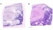
Fluorescence confocal microscopy for evaluation of fresh surgical specimens and consecutive tumor cell isolation in rare pediatric tumors.
Fluorescence confocal microscopy (FCM) is an optical technique that uses laser light sources of different wavelengths to generate real-time images of fresh, unfixed tissue specimens. FCM allows histological evaluation of fresh tissue samples without the associated cryo artifacts after frozen sectioning. The aim of this study was to prospectively evaluate pediatric tumor specimens and assess their suitability for fresh tumor sampling. In addition, we aimed to determine whether tumor cell isolation for stable cell culture is still feasible after FCM imaging. Pediatric tumor specimens were imaged using FCM. Tumor viability and suitability for tissue sampling were evaluated and compared with H&E staining after paraffin embedding. In addition, FCM-processed and non-FCM-processed tissue samples were sent for tumor cell isolation to evaluate possible effects after FCM processing. When comparing estimated tumor cell viability using FCM and H&E, we found good to excellent correlating estimates (intraclass correlation coefficient = 0.891, p < 0.001), as well as substantial agreement in whether the tissue appeared adequate for fresh tissue collection (κ = 0.762, p < 0.001). After FCM, seven out of eight samples yielded passable cell cultures, compared to eight out of eight for non-FCM processed samples. Our study suggests that the use of FCM in tumor sampling can increase the yield of suitable fresh tumor samples by identifying viable tumor areas and ensuring that sufficient tissue remains for diagnosis. Our study also provides first evidence that the isolation and growth of tumor cells in culture are not compromised by the FCM technique.
期刊介绍:
Manuscripts of original studies reinforcing the evidence base of modern diagnostic pathology, using immunocytochemical, molecular and ultrastructural techniques, will be welcomed. In addition, papers on critical evaluation of diagnostic criteria but also broadsheets and guidelines with a solid evidence base will be considered. Consideration will also be given to reports of work in other fields relevant to the understanding of human pathology as well as manuscripts on the application of new methods and techniques in pathology. Submission of purely experimental articles is discouraged but manuscripts on experimental work applicable to diagnostic pathology are welcomed. Biomarker studies are welcomed but need to abide by strict rules (e.g. REMARK) of adequate sample size and relevant marker choice. Single marker studies on limited patient series without validated application will as a rule not be considered. Case reports will only be considered when they provide substantial new information with an impact on understanding disease or diagnostic practice.

 求助内容:
求助内容: 应助结果提醒方式:


