{"title":"坏死深度能否替代坏死体积来预测股骨头坏死的塌陷进展?","authors":"Tomoya Nawata, Takeshi Utsunomiya, Goro Motomura, Ryosuke Yamaguchi, Satoshi Hamai, Shinya Kawahara, Taishi Sato, Daisuke Hara, Kenji Kitamura, Yasuharu Nakashima","doi":"10.1007/s00256-024-04741-0","DOIUrl":null,"url":null,"abstract":"<p><strong>Objective: </strong>Although some patients may experience collapse progression while others may not in post-collapse osteonecrosis of the femoral head (ONFH) with a necrotic lesion located within the weight-bearing part of the acetabulum (Type B/C1), few studies have focused on the natural course after collapse. This study aimed to clarify the correlation between necrotic volume (NV) and necrotic depth (ND) in predicting collapse progression in patients with post-collapse ONFH Type B/C1.</p><p><strong>Materials and methods: </strong>We retrospectively reviewed 54 hips with post-collapse ONFH Type B/C1 from 52 consecutive patients who were conservatively followed up for more than 1 year. We measured the amount of femoral head collapse using biplane radiographs at each follow-up period, and produced Kaplan-Meier survival curves with collapse progression (≥ 1 mm) as the endpoint. We compared NV and ND, which were calculated as the ratio of the distance from the articular surface of the femoral head to the deepest point of a necrotic lesion to the femoral head diameter in the mid-coronal slice of T1-weighted magnetic resonance imaging (MRI).</p><p><strong>Results: </strong>We observed collapse progression in 31 hips (57.4%). The NV and ND were significantly greater in hips with collapse progression than in those without collapse progression (p = 0.0127 and 0.0047, respectively). Necrotic volume was significantly correlated with ND (rs = 0.56, p < 0.0001).</p><p><strong>Conclusion: </strong>This study suggests that necrotic depth on the mid-coronal slice of T1-weighted MRI can be a substitute for necrotic volume to predict collapse progression in ONFH Type B/C1.</p>","PeriodicalId":21783,"journal":{"name":"Skeletal Radiology","volume":" ","pages":"317-324"},"PeriodicalIF":1.9000,"publicationDate":"2025-02-01","publicationTypes":"Journal Article","fieldsOfStudy":null,"isOpenAccess":false,"openAccessPdf":"","citationCount":"0","resultStr":"{\"title\":\"Can necrotic depth be a substitute of necrotic volume to predict collapse progression in osteonecrosis of the femoral head?\",\"authors\":\"Tomoya Nawata, Takeshi Utsunomiya, Goro Motomura, Ryosuke Yamaguchi, Satoshi Hamai, Shinya Kawahara, Taishi Sato, Daisuke Hara, Kenji Kitamura, Yasuharu Nakashima\",\"doi\":\"10.1007/s00256-024-04741-0\",\"DOIUrl\":null,\"url\":null,\"abstract\":\"<p><strong>Objective: </strong>Although some patients may experience collapse progression while others may not in post-collapse osteonecrosis of the femoral head (ONFH) with a necrotic lesion located within the weight-bearing part of the acetabulum (Type B/C1), few studies have focused on the natural course after collapse. This study aimed to clarify the correlation between necrotic volume (NV) and necrotic depth (ND) in predicting collapse progression in patients with post-collapse ONFH Type B/C1.</p><p><strong>Materials and methods: </strong>We retrospectively reviewed 54 hips with post-collapse ONFH Type B/C1 from 52 consecutive patients who were conservatively followed up for more than 1 year. We measured the amount of femoral head collapse using biplane radiographs at each follow-up period, and produced Kaplan-Meier survival curves with collapse progression (≥ 1 mm) as the endpoint. We compared NV and ND, which were calculated as the ratio of the distance from the articular surface of the femoral head to the deepest point of a necrotic lesion to the femoral head diameter in the mid-coronal slice of T1-weighted magnetic resonance imaging (MRI).</p><p><strong>Results: </strong>We observed collapse progression in 31 hips (57.4%). The NV and ND were significantly greater in hips with collapse progression than in those without collapse progression (p = 0.0127 and 0.0047, respectively). Necrotic volume was significantly correlated with ND (rs = 0.56, p < 0.0001).</p><p><strong>Conclusion: </strong>This study suggests that necrotic depth on the mid-coronal slice of T1-weighted MRI can be a substitute for necrotic volume to predict collapse progression in ONFH Type B/C1.</p>\",\"PeriodicalId\":21783,\"journal\":{\"name\":\"Skeletal Radiology\",\"volume\":\" \",\"pages\":\"317-324\"},\"PeriodicalIF\":1.9000,\"publicationDate\":\"2025-02-01\",\"publicationTypes\":\"Journal Article\",\"fieldsOfStudy\":null,\"isOpenAccess\":false,\"openAccessPdf\":\"\",\"citationCount\":\"0\",\"resultStr\":null,\"platform\":\"Semanticscholar\",\"paperid\":null,\"PeriodicalName\":\"Skeletal Radiology\",\"FirstCategoryId\":\"3\",\"ListUrlMain\":\"https://doi.org/10.1007/s00256-024-04741-0\",\"RegionNum\":3,\"RegionCategory\":\"医学\",\"ArticlePicture\":[],\"TitleCN\":null,\"AbstractTextCN\":null,\"PMCID\":null,\"EPubDate\":\"2024/7/9 0:00:00\",\"PubModel\":\"Epub\",\"JCR\":\"Q2\",\"JCRName\":\"ORTHOPEDICS\",\"Score\":null,\"Total\":0}","platform":"Semanticscholar","paperid":null,"PeriodicalName":"Skeletal Radiology","FirstCategoryId":"3","ListUrlMain":"https://doi.org/10.1007/s00256-024-04741-0","RegionNum":3,"RegionCategory":"医学","ArticlePicture":[],"TitleCN":null,"AbstractTextCN":null,"PMCID":null,"EPubDate":"2024/7/9 0:00:00","PubModel":"Epub","JCR":"Q2","JCRName":"ORTHOPEDICS","Score":null,"Total":0}
Can necrotic depth be a substitute of necrotic volume to predict collapse progression in osteonecrosis of the femoral head?
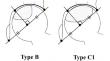
Objective: Although some patients may experience collapse progression while others may not in post-collapse osteonecrosis of the femoral head (ONFH) with a necrotic lesion located within the weight-bearing part of the acetabulum (Type B/C1), few studies have focused on the natural course after collapse. This study aimed to clarify the correlation between necrotic volume (NV) and necrotic depth (ND) in predicting collapse progression in patients with post-collapse ONFH Type B/C1.
Materials and methods: We retrospectively reviewed 54 hips with post-collapse ONFH Type B/C1 from 52 consecutive patients who were conservatively followed up for more than 1 year. We measured the amount of femoral head collapse using biplane radiographs at each follow-up period, and produced Kaplan-Meier survival curves with collapse progression (≥ 1 mm) as the endpoint. We compared NV and ND, which were calculated as the ratio of the distance from the articular surface of the femoral head to the deepest point of a necrotic lesion to the femoral head diameter in the mid-coronal slice of T1-weighted magnetic resonance imaging (MRI).
Results: We observed collapse progression in 31 hips (57.4%). The NV and ND were significantly greater in hips with collapse progression than in those without collapse progression (p = 0.0127 and 0.0047, respectively). Necrotic volume was significantly correlated with ND (rs = 0.56, p < 0.0001).
Conclusion: This study suggests that necrotic depth on the mid-coronal slice of T1-weighted MRI can be a substitute for necrotic volume to predict collapse progression in ONFH Type B/C1.
期刊介绍:
Skeletal Radiology provides a forum for the dissemination of current knowledge and information dealing with disorders of the musculoskeletal system including the spine. While emphasizing the radiological aspects of the many varied skeletal abnormalities, the journal also adopts an interdisciplinary approach, reflecting the membership of the International Skeletal Society. Thus, the anatomical, pathological, physiological, clinical, metabolic and epidemiological aspects of the many entities affecting the skeleton receive appropriate consideration.
This is the Journal of the International Skeletal Society and the Official Journal of the Society of Skeletal Radiology and the Australasian Musculoskelelal Imaging Group.

 求助内容:
求助内容: 应助结果提醒方式:


