{"title":"大鼠颈动脉窦花喷神经末梢的三维超微结构","authors":"Yusuke Murakami, Kuniaki Sasaki, Misaki Komuro, Takuya Yokoyama, Sayed Sharif Abdali, Nobuaki Nakamuta, Yoshio Yamamoto","doi":"10.1002/cne.25654","DOIUrl":null,"url":null,"abstract":"<p>The flower-spray nerve endings are afferent nerve terminals in the carotid sinus that arise from carotid sinus nerve of glossopharyngeal nerve. However, the three-dimensional ultrastructural characteristics of flower-spray nerve endings and spatial relationships between the terminal parts and other cellular elements have not been fully understood. To elucidate their detailed relationship, backscattered electron imaging of serial sections was performed with a scanning electron microscope to produce a three-dimensional reconstruction of the flower-spray endings. The terminal parts of flower-spray endings were distributed horizontally approximately 5 µm outside the external elastic membrane in the tunica adventitia of the internal carotid artery. The three-dimensional reconstruction showed that the terminal parts of flower-spray endings were flat with irregular contours and were partially covered by the thin cytoplasmic processes of Schwann cells. The complex consisting of the nerve terminals and associated Schwann cells was surrounded by a multilayered basement membrane. The terminal parts of the endings were also surrounded by fibroblasts with elastic fibers and collagen fibrils. Secretory vesicles without an electron-dense core were observed in the terminal parts of the endings. The accumulation of vesicles just below the axonal membrane was observed in terminal parts not covered by Schwann cell cytoplasmic processes on both the luminal and basal sides. Swollen mitochondria, concentric membranous structures, and glycogen granule-like electron-dense materials were often noted in some of the terminal parts of the endings and the parent axon. Collectively, the present results suggest that flower-spray endings are baroreceptors because their morphology was similar to other mechanoreceptors. Furthermore, flower-spray endings may be affected by glutamate secreted in an autocrine manner.</p>","PeriodicalId":15552,"journal":{"name":"Journal of Comparative Neurology","volume":"532 7","pages":""},"PeriodicalIF":2.3000,"publicationDate":"2024-07-09","publicationTypes":"Journal Article","fieldsOfStudy":null,"isOpenAccess":false,"openAccessPdf":"https://onlinelibrary.wiley.com/doi/epdf/10.1002/cne.25654","citationCount":"0","resultStr":"{\"title\":\"Three-Dimensional Ultrastructure of Flower-Spray Nerve Endings in the Rat Carotid Sinus\",\"authors\":\"Yusuke Murakami, Kuniaki Sasaki, Misaki Komuro, Takuya Yokoyama, Sayed Sharif Abdali, Nobuaki Nakamuta, Yoshio Yamamoto\",\"doi\":\"10.1002/cne.25654\",\"DOIUrl\":null,\"url\":null,\"abstract\":\"<p>The flower-spray nerve endings are afferent nerve terminals in the carotid sinus that arise from carotid sinus nerve of glossopharyngeal nerve. However, the three-dimensional ultrastructural characteristics of flower-spray nerve endings and spatial relationships between the terminal parts and other cellular elements have not been fully understood. To elucidate their detailed relationship, backscattered electron imaging of serial sections was performed with a scanning electron microscope to produce a three-dimensional reconstruction of the flower-spray endings. The terminal parts of flower-spray endings were distributed horizontally approximately 5 µm outside the external elastic membrane in the tunica adventitia of the internal carotid artery. The three-dimensional reconstruction showed that the terminal parts of flower-spray endings were flat with irregular contours and were partially covered by the thin cytoplasmic processes of Schwann cells. The complex consisting of the nerve terminals and associated Schwann cells was surrounded by a multilayered basement membrane. The terminal parts of the endings were also surrounded by fibroblasts with elastic fibers and collagen fibrils. Secretory vesicles without an electron-dense core were observed in the terminal parts of the endings. The accumulation of vesicles just below the axonal membrane was observed in terminal parts not covered by Schwann cell cytoplasmic processes on both the luminal and basal sides. Swollen mitochondria, concentric membranous structures, and glycogen granule-like electron-dense materials were often noted in some of the terminal parts of the endings and the parent axon. Collectively, the present results suggest that flower-spray endings are baroreceptors because their morphology was similar to other mechanoreceptors. Furthermore, flower-spray endings may be affected by glutamate secreted in an autocrine manner.</p>\",\"PeriodicalId\":15552,\"journal\":{\"name\":\"Journal of Comparative Neurology\",\"volume\":\"532 7\",\"pages\":\"\"},\"PeriodicalIF\":2.3000,\"publicationDate\":\"2024-07-09\",\"publicationTypes\":\"Journal Article\",\"fieldsOfStudy\":null,\"isOpenAccess\":false,\"openAccessPdf\":\"https://onlinelibrary.wiley.com/doi/epdf/10.1002/cne.25654\",\"citationCount\":\"0\",\"resultStr\":null,\"platform\":\"Semanticscholar\",\"paperid\":null,\"PeriodicalName\":\"Journal of Comparative Neurology\",\"FirstCategoryId\":\"3\",\"ListUrlMain\":\"https://onlinelibrary.wiley.com/doi/10.1002/cne.25654\",\"RegionNum\":4,\"RegionCategory\":\"医学\",\"ArticlePicture\":[],\"TitleCN\":null,\"AbstractTextCN\":null,\"PMCID\":null,\"EPubDate\":\"\",\"PubModel\":\"\",\"JCR\":\"Q3\",\"JCRName\":\"NEUROSCIENCES\",\"Score\":null,\"Total\":0}","platform":"Semanticscholar","paperid":null,"PeriodicalName":"Journal of Comparative Neurology","FirstCategoryId":"3","ListUrlMain":"https://onlinelibrary.wiley.com/doi/10.1002/cne.25654","RegionNum":4,"RegionCategory":"医学","ArticlePicture":[],"TitleCN":null,"AbstractTextCN":null,"PMCID":null,"EPubDate":"","PubModel":"","JCR":"Q3","JCRName":"NEUROSCIENCES","Score":null,"Total":0}
Three-Dimensional Ultrastructure of Flower-Spray Nerve Endings in the Rat Carotid Sinus
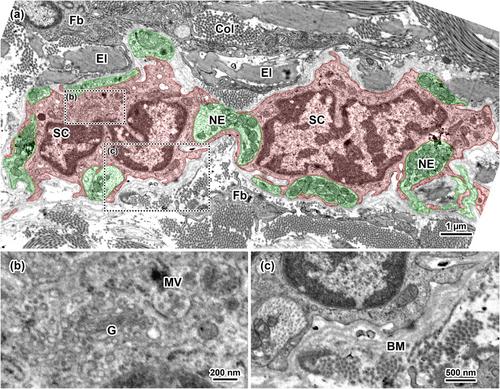
The flower-spray nerve endings are afferent nerve terminals in the carotid sinus that arise from carotid sinus nerve of glossopharyngeal nerve. However, the three-dimensional ultrastructural characteristics of flower-spray nerve endings and spatial relationships between the terminal parts and other cellular elements have not been fully understood. To elucidate their detailed relationship, backscattered electron imaging of serial sections was performed with a scanning electron microscope to produce a three-dimensional reconstruction of the flower-spray endings. The terminal parts of flower-spray endings were distributed horizontally approximately 5 µm outside the external elastic membrane in the tunica adventitia of the internal carotid artery. The three-dimensional reconstruction showed that the terminal parts of flower-spray endings were flat with irregular contours and were partially covered by the thin cytoplasmic processes of Schwann cells. The complex consisting of the nerve terminals and associated Schwann cells was surrounded by a multilayered basement membrane. The terminal parts of the endings were also surrounded by fibroblasts with elastic fibers and collagen fibrils. Secretory vesicles without an electron-dense core were observed in the terminal parts of the endings. The accumulation of vesicles just below the axonal membrane was observed in terminal parts not covered by Schwann cell cytoplasmic processes on both the luminal and basal sides. Swollen mitochondria, concentric membranous structures, and glycogen granule-like electron-dense materials were often noted in some of the terminal parts of the endings and the parent axon. Collectively, the present results suggest that flower-spray endings are baroreceptors because their morphology was similar to other mechanoreceptors. Furthermore, flower-spray endings may be affected by glutamate secreted in an autocrine manner.
期刊介绍:
Established in 1891, JCN is the oldest continually published basic neuroscience journal. Historically, as the name suggests, the journal focused on a comparison among species to uncover the intricacies of how the brain functions. In modern times, this research is called systems neuroscience where animal models are used to mimic core cognitive processes with the ultimate goal of understanding neural circuits and connections that give rise to behavioral patterns and different neural states.
Research published in JCN covers all species from invertebrates to humans, and the reports inform the readers about the function and organization of nervous systems in species with an emphasis on the way that species adaptations inform about the function or organization of the nervous systems, rather than on their evolution per se.
JCN publishes primary research articles and critical commentaries and review-type articles offering expert insight in to cutting edge research in the field of systems neuroscience; a complete list of contribution types is given in the Author Guidelines. For primary research contributions, only full-length investigative reports are desired; the journal does not accept short communications.

 求助内容:
求助内容: 应助结果提醒方式:


