Michael Schnekenburger, Anne Lorant, Sruthi Reddy Gajulapalli, Ridhika Rajora, Jin-Young Lee, Aloran Mazumder, Haeun Yang, Christo Christov, Hyoung Jin Kang, Bernard Pirotte, Marc Diederich
{"title":"双重抑制 sirtuins 1 和 2:重新规划慢性髓性白血病的代谢能量动态,作为一种免疫原性抗癌策略。","authors":"Michael Schnekenburger, Anne Lorant, Sruthi Reddy Gajulapalli, Ridhika Rajora, Jin-Young Lee, Aloran Mazumder, Haeun Yang, Christo Christov, Hyoung Jin Kang, Bernard Pirotte, Marc Diederich","doi":"10.1002/cac2.12590","DOIUrl":null,"url":null,"abstract":"<p>Chronic myeloid leukemia (CML) is a lethal hematopoietic malignancy with a global incidence primarily attributed to the <i>breakpoint cluster region-Abelson</i> (BCR-ABL1) fusion oncogene in over 95% of cases. The introduction of tyrosine kinase inhibitors (TKIs) has revolutionized CML management; however, a subset of patients encounters challenges such as resistance and relapse, hindering the achievement of complete remission. Overcoming these challenges in CML also requires addressing persistent leukemia stem cells (LSCs) with inherent resistance mechanisms. Key regulators of LSC metabolism, proliferation, and survival, as well as genetic and epigenetic alterations, provide potential targets [<span>1</span>].</p><p>In this study, leveraging in silico analysis (methods and descriptions of other assays are in the Supplementary file) of LSCs from CML patients at diagnosis, we demonstrated enrichment in pathways predominantly associated with proliferation, oxidative phosphorylation (OXPHOS), and metabolism, concurrently with a decrease in immune response pathways (Figure 1A). Similarly, genes negatively impacting proliferation in CML cell lines, when depleted by CRISPR, were enriched in processes related to OXPHOS, metabolism, and proliferation, mirroring the enrichment observed in LSCs (Figure 1B) [<span>2</span>].</p><p>Sirtuins (SIRTs) are nicotinamide adenine dinucleotide (NAD)<sup>+</sup>-dependent histone deacetylases. SIRT1 and SIRT2 modulate key signaling proteins impacting metabolism, survival, and stress response [<span>3</span>]. Overexpression of SIRT1 and SIRT2 was observed in various cancers, including leukemia (Supplementary Figure S1A) [<span>4, 5</span>]. However, our analysis of CML patients revealed variability in the expression levels of SIRT1 and SIRT2 across datasets (Supplementary Figure S1B). Given the relatively small sample sizes, we recognized the limitations of relying solely on single gene expression data, as it may not fully capture the functional relevance of SIRT1/2 in CML. In response to this limitation, we expanded our analysis to identify broader gene expression patterns associated with SIRT1/2. Specifically, we identified a CML-related network comprising 180 co-regulated transcriptional targets associated with SIRT1/2 enriched in genes relevant to leukemia (Figure 1C, Supplementary Figure S1C-E). To quantify their collective impact, we consolidated the expression of all transcripts in the SIRT1/2 regulon into a unified score, referred to as the SIRT1-2 regulon score. This score effectively discriminated between healthy hematopoietic stem cells and LSCs from CML patients at diagnosis (Figure 1D), indicating the collective impact of SIRT1 and SIRT2 on the disease.</p><p>Given the complementary roles of SIRT1 and 2 in regulating metabolic and survival pathways and their potential to compensate for each other's loss of function [<span>3</span>], we postulated that exploiting metabolic vulnerabilities in LSCs through dual targeting of SIRT1 and SIRT2 activities could profoundly influence CML cell survival. We first verified the constitutive SIRT1 and SIRT2 expression levels in myeloid cell lines and showed that their genetic and pharmacological inhibition, including with the compound Si-711 [<span>6</span>], reduced proliferation and total HDAC activities in CML cells (Supplementary Figures S1F, S2-S3). Si-711 has favorable pharmacokinetics and pharmacodynamics parameters and demonstrated superior activities compared to other reference SIRT inhibitors (Supplementary Tables S1-S3,Supplementary Figures S4-S5). We demonstrated that Si-711 induced robust cytostatic effects concomitant with G1 arrest in various myeloid leukemia cells, including both TKI-sensitive and -resistant models, exhibiting a good selectivity for cancer cells (selectivity factor ranging from 3 to 18) compared to normal cells (Supplementary Table S3, Supplementary Figures S6-S7, S8A). Inhibiting SIRT1/2 activities reduced the viability of primary CML cells from patients <i>ex vivo</i> and significantly inhibited cell growth in leukemia patient-derived xenograft zebrafish and CML cell xenograft mouse models without signs of toxicity (Figure 1E-F, Supplementary Figure S9, Supplementary Tables S4-S5). Additionally, Si-711 treatment significantly reduced the aldehyde dehydrogenase-positive subpopulation of CML cells (Supplementary Figure S10), a marker of stemness, indicating its potential efficacy against LSCs. Moreover, Si-711 strongly synergized with imatinib, the first-line therapy against CML, in both imatinib-sensitive and -resistant CML cell lines (Figure 1G, Supplementary Figure S11), potentially overcoming resistance mechanisms observed in some CML cases.</p><p>RNA-sequencing of Si-711-treated K-562 CML cells for up to 48 h was conducted to investigate mechanistic aspects. We validated the SIRT1-2 score established from patient data, showing that Si-711 significantly reduced this score in a time-dependent manner (Pearson test, <i>r =</i> 0.92, <i>P</i> < 0.001; Figure 1H, Supplementary Figure S12A). Differentially expressed genes were analyzed, revealing a downregulation of genes involved in proliferation, enriched in those regulated by key transcription factors such as MYC, JUN, and E2F4, which have extensive connections to the SIRT1/2 regulon, and an upregulation of genes associated with the immune response and cell death mechanisms (Figure 1I, Supplementary Figures S12B-H, S13). Metabolic reaction enrichment analysis revealed that, at 24 h, Si-711 induced significant metabolic alterations in CML cells, including upregulation of glycolysis and downregulation of nucleotide metabolism and mitochondrial respiratory complexes (Supplementary Figure S12I-Q). <i>In cellulo</i> investigations revealed that SIRT1/2 inhibition induced critical metabolic alterations in CML, leading to a decrease in oxygen consumption rate (OCR), a transient increase in extracellular acidification rate (ECAR), and ultimately, a reduction in overall metabolic activity (Figure 1J, Supplementary Figure S14A-G). As an adaptive response to the inhibition of OCR, Si-711 enhanced glycolysis, leading to increased extracellular acidification, lactate release, and a shift of CML cells towards predominant glycolytic-dependent ATP production, deviating from their initial reliance on OXPHOS-dependent ATP (Figure 1K-M, Supplementary Figures S8B, S14H-K, S15A), a characteristic of LSCs and therapy-resistant cells [<span>7</span>]. Ultimately, these metabolic alterations resulted in a progressive depletion in total ATP content, significant changes in the NAD<sup>+</sup>/NADH ratio, and morphological alterations in mitochondria, including onion-like swirling cores surrounded by swollen spaces, accompanied by a less condensed and disorganized cristae structure (Figure 1N, Supplementary Figures S8C, S14L, S15B-C, S16-S18). Furthermore, Si-711 treatment increased mitochondrial superoxide levels, suggesting the induction of oxidative stress (Figure 1O, Supplementary Figure S19). Upon prolonged exposure, the resulting energetic collapse directed the fate of most myeloid leukemia models toward necrosis-like cell death at concentrations ranging from 5-10 µmol/L, selectively targeting cancer cells with selectivity factors ranging from 10 to 105 (Figure 1P, Supplementary Figures S8D, S20-S22, Supplementary Table S6). This non-canonical cell death is distinct from traditional anti-CML therapies, holding promise for overcoming canonical apoptotic resistance mechanisms [<span>8</span>]. The inhibition of SIRT1/2 induced a caspase/RIPK3-independent but RIPK1/MLKL/PARP1-dependent necrosis (Figure 1Q, Supplementary Figures S23-S26). This necrosis led to a progressive depletion of endoplasmic reticulum calcium storage, causing its accumulation in mitochondria associated with the opening of the mitochondrial permeability transition pore (Supplementary Figures S24, S27-S30). SIRT1/2 inhibition with Si-711 induced immunologic regulated necrosis in CML cells, significantly promoting the release of damage-associated molecular patterns (DAMPs), including calreticulin, high mobility group box 1 (HMGB1), and ATP (Figure 1R-U, Supplementary Figure S31A-E). This can potentially trigger immunogenic cell death (ICD) and enhance the anticancer immune response [<span>9</span>]. Notably, Si-711 treatment boosted phagocytosis (Figure 1V, Supplementary Figure S31F), indicating a potential increase in immune response. We conducted an in vivo vaccination assay, which is considered the gold standard to validate the induction of ICD and assess therapeutic potential [<span>10</span>]. Upon tumor challenge, mice vaccinated with Si-711-treated cells exhibited a significant reduction in tumor volume, tumor weight, and splenomegaly (Figure 1W, Supplementary Figures S32-S33). Importantly, Si-711's effects in inducing ICD were found to be comparable to the known ICD inducer oxaliplatin.</p><p>In conclusion, we propose SIRT1/2 inhibition, particularly with Si-711, as a promising and innovative therapeutic strategy for CML. The dual targeting of SIRT1 and SIRT2 could eradicate LSCs through metabolic reprogramming and induction of immunogenic necrosis-like cell death. This comprehensive approach holds the potential for addressing challenges such as resistance and relapse in CML, providing new avenues for improved patient outcomes.</p><p>Michael Schnekenburger performed in vitro/<i>in cellulo</i> experiments; Sruthi Reddy Gajulapalli, Ridhika Rajora, and Jin-Young Lee performed in vivo experiments; Hyoung Jin Kang provided patient samples; Aloran Mazumder and Haeun Yang performed additional in vitro experiments; Anne Lorant performed, analyzed, and interpreted bioinformatic data and statistical analyses; Christo Christov interpreted and analyzed histological sections; Michael Schnekenburger and Marc Diederich conceived and designed the project. Michael Schnekenburger, Anne Lorant, and Marc Diederich wrote/edited the manuscript; Bernard Pirotte synthesized the inhibitor; Marc Diederich supervised the project. All authors have read and approved the manuscript. Marc Diederich is responsible for all aspects of this work, including descriptions, illustrations, and data accuracy.</p><p>The authors declare no conflict of interest.</p><p>LBMCC: “Recherche Cancer et Sang” foundation, the “Recherches Scientifiques Luxembourg”, the “Een Häerz fir kriibskrank Kanner”, the Action LIONS “Vaincre le Cancer” and Télévie Luxembourg. SNU: National Research Foundation (NRF) (Grant Number 370C-20220063); MEST of Korea for Tumor Microenvironment Global Core Research Center (GCRC) (Grant Number 2011-0030001); Brain Korea (BK21) PLUS program and Creative-Pioneering Researchers Program at Seoul National University (Funding number: 370C-20160062). Sruthi Reddy Gajulapalli and Ridhika Rajora were supported by grants for the “Graduate Scholarship for Excellent Foreign Students” program.</p><p>The data generated during this study are included in this published article and its supplementary information files.</p><p>The datasets analyzed during the current study are available from the corresponding author upon reasonable request.</p><p>All animal experiments were conducted following the guidelines set by the “Korean Food and Drug Administration”. Protocols were reviewed and approved by the “Institutional Animal Care and Use Committee of Seoul National University” (approval numbers: SNU-201006-2 [NOD/SCID xenograft model], SNU-210518-10-2 [vaccination assay], SNU-191218-5-1 [zebrafish]).</p><p>For human leukemia samples, the Institutional Review Board of Seoul National University Hospital reviewed and approved the study protocol and exempted the study from the obligation to obtain informed consent. This study was performed following the World Medical Association's <b>Declaration of Helsinki</b>.</p><p>PBMCs and P_PBMCs were used with the approval of the National Research Ethics Committee of Luxembourg. PBMCs were isolated from blood obtained from the Red Cross (Luxembourg, Luxembourg) under the authorization LBMCC-2019-0002: “Assessment of toxicity of new drugs or drug combinations in preclinical development in nonproliferating peripheral blood mononuclear cells (systemic acute toxicity)”. P_PBMCs were generated from blood obtained from the Red Cross under the authorization LBMCC- 2019-0001: “Assessment of differential toxicity of new drugs or drug combinations in preclinical development in <i>ex-vivo</i> proliferating peripheral blood mononuclear cells vs. proliferating cancer cells.”</p><p>The obtention of umbilical cord blood samples for the isolation of CD34<sup>+</sup> cells was approved by the board of directors of the Bohler clinic (Hôpitaux Robert Schuman, Luxembourg, Luxembourg). Tissues were obtained anonymously with the written informed consent of parents with the approval of the National Research Ethics Committee of Luxembourg.</p>","PeriodicalId":9495,"journal":{"name":"Cancer Communications","volume":"44 8","pages":"915-920"},"PeriodicalIF":20.1000,"publicationDate":"2024-07-08","publicationTypes":"Journal Article","fieldsOfStudy":null,"isOpenAccess":false,"openAccessPdf":"https://onlinelibrary.wiley.com/doi/epdf/10.1002/cac2.12590","citationCount":"0","resultStr":"{\"title\":\"Dual inhibition of sirtuins 1 and 2: reprogramming metabolic energy dynamics in chronic myeloid leukemia as an immunogenic anticancer strategy\",\"authors\":\"Michael Schnekenburger, Anne Lorant, Sruthi Reddy Gajulapalli, Ridhika Rajora, Jin-Young Lee, Aloran Mazumder, Haeun Yang, Christo Christov, Hyoung Jin Kang, Bernard Pirotte, Marc Diederich\",\"doi\":\"10.1002/cac2.12590\",\"DOIUrl\":null,\"url\":null,\"abstract\":\"<p>Chronic myeloid leukemia (CML) is a lethal hematopoietic malignancy with a global incidence primarily attributed to the <i>breakpoint cluster region-Abelson</i> (BCR-ABL1) fusion oncogene in over 95% of cases. The introduction of tyrosine kinase inhibitors (TKIs) has revolutionized CML management; however, a subset of patients encounters challenges such as resistance and relapse, hindering the achievement of complete remission. Overcoming these challenges in CML also requires addressing persistent leukemia stem cells (LSCs) with inherent resistance mechanisms. Key regulators of LSC metabolism, proliferation, and survival, as well as genetic and epigenetic alterations, provide potential targets [<span>1</span>].</p><p>In this study, leveraging in silico analysis (methods and descriptions of other assays are in the Supplementary file) of LSCs from CML patients at diagnosis, we demonstrated enrichment in pathways predominantly associated with proliferation, oxidative phosphorylation (OXPHOS), and metabolism, concurrently with a decrease in immune response pathways (Figure 1A). Similarly, genes negatively impacting proliferation in CML cell lines, when depleted by CRISPR, were enriched in processes related to OXPHOS, metabolism, and proliferation, mirroring the enrichment observed in LSCs (Figure 1B) [<span>2</span>].</p><p>Sirtuins (SIRTs) are nicotinamide adenine dinucleotide (NAD)<sup>+</sup>-dependent histone deacetylases. SIRT1 and SIRT2 modulate key signaling proteins impacting metabolism, survival, and stress response [<span>3</span>]. Overexpression of SIRT1 and SIRT2 was observed in various cancers, including leukemia (Supplementary Figure S1A) [<span>4, 5</span>]. However, our analysis of CML patients revealed variability in the expression levels of SIRT1 and SIRT2 across datasets (Supplementary Figure S1B). Given the relatively small sample sizes, we recognized the limitations of relying solely on single gene expression data, as it may not fully capture the functional relevance of SIRT1/2 in CML. In response to this limitation, we expanded our analysis to identify broader gene expression patterns associated with SIRT1/2. Specifically, we identified a CML-related network comprising 180 co-regulated transcriptional targets associated with SIRT1/2 enriched in genes relevant to leukemia (Figure 1C, Supplementary Figure S1C-E). To quantify their collective impact, we consolidated the expression of all transcripts in the SIRT1/2 regulon into a unified score, referred to as the SIRT1-2 regulon score. This score effectively discriminated between healthy hematopoietic stem cells and LSCs from CML patients at diagnosis (Figure 1D), indicating the collective impact of SIRT1 and SIRT2 on the disease.</p><p>Given the complementary roles of SIRT1 and 2 in regulating metabolic and survival pathways and their potential to compensate for each other's loss of function [<span>3</span>], we postulated that exploiting metabolic vulnerabilities in LSCs through dual targeting of SIRT1 and SIRT2 activities could profoundly influence CML cell survival. We first verified the constitutive SIRT1 and SIRT2 expression levels in myeloid cell lines and showed that their genetic and pharmacological inhibition, including with the compound Si-711 [<span>6</span>], reduced proliferation and total HDAC activities in CML cells (Supplementary Figures S1F, S2-S3). Si-711 has favorable pharmacokinetics and pharmacodynamics parameters and demonstrated superior activities compared to other reference SIRT inhibitors (Supplementary Tables S1-S3,Supplementary Figures S4-S5). We demonstrated that Si-711 induced robust cytostatic effects concomitant with G1 arrest in various myeloid leukemia cells, including both TKI-sensitive and -resistant models, exhibiting a good selectivity for cancer cells (selectivity factor ranging from 3 to 18) compared to normal cells (Supplementary Table S3, Supplementary Figures S6-S7, S8A). Inhibiting SIRT1/2 activities reduced the viability of primary CML cells from patients <i>ex vivo</i> and significantly inhibited cell growth in leukemia patient-derived xenograft zebrafish and CML cell xenograft mouse models without signs of toxicity (Figure 1E-F, Supplementary Figure S9, Supplementary Tables S4-S5). Additionally, Si-711 treatment significantly reduced the aldehyde dehydrogenase-positive subpopulation of CML cells (Supplementary Figure S10), a marker of stemness, indicating its potential efficacy against LSCs. Moreover, Si-711 strongly synergized with imatinib, the first-line therapy against CML, in both imatinib-sensitive and -resistant CML cell lines (Figure 1G, Supplementary Figure S11), potentially overcoming resistance mechanisms observed in some CML cases.</p><p>RNA-sequencing of Si-711-treated K-562 CML cells for up to 48 h was conducted to investigate mechanistic aspects. We validated the SIRT1-2 score established from patient data, showing that Si-711 significantly reduced this score in a time-dependent manner (Pearson test, <i>r =</i> 0.92, <i>P</i> < 0.001; Figure 1H, Supplementary Figure S12A). Differentially expressed genes were analyzed, revealing a downregulation of genes involved in proliferation, enriched in those regulated by key transcription factors such as MYC, JUN, and E2F4, which have extensive connections to the SIRT1/2 regulon, and an upregulation of genes associated with the immune response and cell death mechanisms (Figure 1I, Supplementary Figures S12B-H, S13). Metabolic reaction enrichment analysis revealed that, at 24 h, Si-711 induced significant metabolic alterations in CML cells, including upregulation of glycolysis and downregulation of nucleotide metabolism and mitochondrial respiratory complexes (Supplementary Figure S12I-Q). <i>In cellulo</i> investigations revealed that SIRT1/2 inhibition induced critical metabolic alterations in CML, leading to a decrease in oxygen consumption rate (OCR), a transient increase in extracellular acidification rate (ECAR), and ultimately, a reduction in overall metabolic activity (Figure 1J, Supplementary Figure S14A-G). As an adaptive response to the inhibition of OCR, Si-711 enhanced glycolysis, leading to increased extracellular acidification, lactate release, and a shift of CML cells towards predominant glycolytic-dependent ATP production, deviating from their initial reliance on OXPHOS-dependent ATP (Figure 1K-M, Supplementary Figures S8B, S14H-K, S15A), a characteristic of LSCs and therapy-resistant cells [<span>7</span>]. Ultimately, these metabolic alterations resulted in a progressive depletion in total ATP content, significant changes in the NAD<sup>+</sup>/NADH ratio, and morphological alterations in mitochondria, including onion-like swirling cores surrounded by swollen spaces, accompanied by a less condensed and disorganized cristae structure (Figure 1N, Supplementary Figures S8C, S14L, S15B-C, S16-S18). Furthermore, Si-711 treatment increased mitochondrial superoxide levels, suggesting the induction of oxidative stress (Figure 1O, Supplementary Figure S19). Upon prolonged exposure, the resulting energetic collapse directed the fate of most myeloid leukemia models toward necrosis-like cell death at concentrations ranging from 5-10 µmol/L, selectively targeting cancer cells with selectivity factors ranging from 10 to 105 (Figure 1P, Supplementary Figures S8D, S20-S22, Supplementary Table S6). This non-canonical cell death is distinct from traditional anti-CML therapies, holding promise for overcoming canonical apoptotic resistance mechanisms [<span>8</span>]. The inhibition of SIRT1/2 induced a caspase/RIPK3-independent but RIPK1/MLKL/PARP1-dependent necrosis (Figure 1Q, Supplementary Figures S23-S26). This necrosis led to a progressive depletion of endoplasmic reticulum calcium storage, causing its accumulation in mitochondria associated with the opening of the mitochondrial permeability transition pore (Supplementary Figures S24, S27-S30). SIRT1/2 inhibition with Si-711 induced immunologic regulated necrosis in CML cells, significantly promoting the release of damage-associated molecular patterns (DAMPs), including calreticulin, high mobility group box 1 (HMGB1), and ATP (Figure 1R-U, Supplementary Figure S31A-E). This can potentially trigger immunogenic cell death (ICD) and enhance the anticancer immune response [<span>9</span>]. Notably, Si-711 treatment boosted phagocytosis (Figure 1V, Supplementary Figure S31F), indicating a potential increase in immune response. We conducted an in vivo vaccination assay, which is considered the gold standard to validate the induction of ICD and assess therapeutic potential [<span>10</span>]. Upon tumor challenge, mice vaccinated with Si-711-treated cells exhibited a significant reduction in tumor volume, tumor weight, and splenomegaly (Figure 1W, Supplementary Figures S32-S33). Importantly, Si-711's effects in inducing ICD were found to be comparable to the known ICD inducer oxaliplatin.</p><p>In conclusion, we propose SIRT1/2 inhibition, particularly with Si-711, as a promising and innovative therapeutic strategy for CML. The dual targeting of SIRT1 and SIRT2 could eradicate LSCs through metabolic reprogramming and induction of immunogenic necrosis-like cell death. This comprehensive approach holds the potential for addressing challenges such as resistance and relapse in CML, providing new avenues for improved patient outcomes.</p><p>Michael Schnekenburger performed in vitro/<i>in cellulo</i> experiments; Sruthi Reddy Gajulapalli, Ridhika Rajora, and Jin-Young Lee performed in vivo experiments; Hyoung Jin Kang provided patient samples; Aloran Mazumder and Haeun Yang performed additional in vitro experiments; Anne Lorant performed, analyzed, and interpreted bioinformatic data and statistical analyses; Christo Christov interpreted and analyzed histological sections; Michael Schnekenburger and Marc Diederich conceived and designed the project. Michael Schnekenburger, Anne Lorant, and Marc Diederich wrote/edited the manuscript; Bernard Pirotte synthesized the inhibitor; Marc Diederich supervised the project. All authors have read and approved the manuscript. Marc Diederich is responsible for all aspects of this work, including descriptions, illustrations, and data accuracy.</p><p>The authors declare no conflict of interest.</p><p>LBMCC: “Recherche Cancer et Sang” foundation, the “Recherches Scientifiques Luxembourg”, the “Een Häerz fir kriibskrank Kanner”, the Action LIONS “Vaincre le Cancer” and Télévie Luxembourg. SNU: National Research Foundation (NRF) (Grant Number 370C-20220063); MEST of Korea for Tumor Microenvironment Global Core Research Center (GCRC) (Grant Number 2011-0030001); Brain Korea (BK21) PLUS program and Creative-Pioneering Researchers Program at Seoul National University (Funding number: 370C-20160062). Sruthi Reddy Gajulapalli and Ridhika Rajora were supported by grants for the “Graduate Scholarship for Excellent Foreign Students” program.</p><p>The data generated during this study are included in this published article and its supplementary information files.</p><p>The datasets analyzed during the current study are available from the corresponding author upon reasonable request.</p><p>All animal experiments were conducted following the guidelines set by the “Korean Food and Drug Administration”. Protocols were reviewed and approved by the “Institutional Animal Care and Use Committee of Seoul National University” (approval numbers: SNU-201006-2 [NOD/SCID xenograft model], SNU-210518-10-2 [vaccination assay], SNU-191218-5-1 [zebrafish]).</p><p>For human leukemia samples, the Institutional Review Board of Seoul National University Hospital reviewed and approved the study protocol and exempted the study from the obligation to obtain informed consent. This study was performed following the World Medical Association's <b>Declaration of Helsinki</b>.</p><p>PBMCs and P_PBMCs were used with the approval of the National Research Ethics Committee of Luxembourg. PBMCs were isolated from blood obtained from the Red Cross (Luxembourg, Luxembourg) under the authorization LBMCC-2019-0002: “Assessment of toxicity of new drugs or drug combinations in preclinical development in nonproliferating peripheral blood mononuclear cells (systemic acute toxicity)”. P_PBMCs were generated from blood obtained from the Red Cross under the authorization LBMCC- 2019-0001: “Assessment of differential toxicity of new drugs or drug combinations in preclinical development in <i>ex-vivo</i> proliferating peripheral blood mononuclear cells vs. proliferating cancer cells.”</p><p>The obtention of umbilical cord blood samples for the isolation of CD34<sup>+</sup> cells was approved by the board of directors of the Bohler clinic (Hôpitaux Robert Schuman, Luxembourg, Luxembourg). Tissues were obtained anonymously with the written informed consent of parents with the approval of the National Research Ethics Committee of Luxembourg.</p>\",\"PeriodicalId\":9495,\"journal\":{\"name\":\"Cancer Communications\",\"volume\":\"44 8\",\"pages\":\"915-920\"},\"PeriodicalIF\":20.1000,\"publicationDate\":\"2024-07-08\",\"publicationTypes\":\"Journal Article\",\"fieldsOfStudy\":null,\"isOpenAccess\":false,\"openAccessPdf\":\"https://onlinelibrary.wiley.com/doi/epdf/10.1002/cac2.12590\",\"citationCount\":\"0\",\"resultStr\":null,\"platform\":\"Semanticscholar\",\"paperid\":null,\"PeriodicalName\":\"Cancer Communications\",\"FirstCategoryId\":\"3\",\"ListUrlMain\":\"https://onlinelibrary.wiley.com/doi/10.1002/cac2.12590\",\"RegionNum\":1,\"RegionCategory\":\"医学\",\"ArticlePicture\":[],\"TitleCN\":null,\"AbstractTextCN\":null,\"PMCID\":null,\"EPubDate\":\"\",\"PubModel\":\"\",\"JCR\":\"Q1\",\"JCRName\":\"ONCOLOGY\",\"Score\":null,\"Total\":0}","platform":"Semanticscholar","paperid":null,"PeriodicalName":"Cancer Communications","FirstCategoryId":"3","ListUrlMain":"https://onlinelibrary.wiley.com/doi/10.1002/cac2.12590","RegionNum":1,"RegionCategory":"医学","ArticlePicture":[],"TitleCN":null,"AbstractTextCN":null,"PMCID":null,"EPubDate":"","PubModel":"","JCR":"Q1","JCRName":"ONCOLOGY","Score":null,"Total":0}
Dual inhibition of sirtuins 1 and 2: reprogramming metabolic energy dynamics in chronic myeloid leukemia as an immunogenic anticancer strategy
Chronic myeloid leukemia (CML) is a lethal hematopoietic malignancy with a global incidence primarily attributed to the breakpoint cluster region-Abelson (BCR-ABL1) fusion oncogene in over 95% of cases. The introduction of tyrosine kinase inhibitors (TKIs) has revolutionized CML management; however, a subset of patients encounters challenges such as resistance and relapse, hindering the achievement of complete remission. Overcoming these challenges in CML also requires addressing persistent leukemia stem cells (LSCs) with inherent resistance mechanisms. Key regulators of LSC metabolism, proliferation, and survival, as well as genetic and epigenetic alterations, provide potential targets [1].
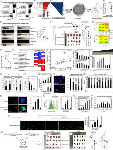
In this study, leveraging in silico analysis (methods and descriptions of other assays are in the Supplementary file) of LSCs from CML patients at diagnosis, we demonstrated enrichment in pathways predominantly associated with proliferation, oxidative phosphorylation (OXPHOS), and metabolism, concurrently with a decrease in immune response pathways (Figure 1A). Similarly, genes negatively impacting proliferation in CML cell lines, when depleted by CRISPR, were enriched in processes related to OXPHOS, metabolism, and proliferation, mirroring the enrichment observed in LSCs (Figure 1B) [2].
Sirtuins (SIRTs) are nicotinamide adenine dinucleotide (NAD)+-dependent histone deacetylases. SIRT1 and SIRT2 modulate key signaling proteins impacting metabolism, survival, and stress response [3]. Overexpression of SIRT1 and SIRT2 was observed in various cancers, including leukemia (Supplementary Figure S1A) [4, 5]. However, our analysis of CML patients revealed variability in the expression levels of SIRT1 and SIRT2 across datasets (Supplementary Figure S1B). Given the relatively small sample sizes, we recognized the limitations of relying solely on single gene expression data, as it may not fully capture the functional relevance of SIRT1/2 in CML. In response to this limitation, we expanded our analysis to identify broader gene expression patterns associated with SIRT1/2. Specifically, we identified a CML-related network comprising 180 co-regulated transcriptional targets associated with SIRT1/2 enriched in genes relevant to leukemia (Figure 1C, Supplementary Figure S1C-E). To quantify their collective impact, we consolidated the expression of all transcripts in the SIRT1/2 regulon into a unified score, referred to as the SIRT1-2 regulon score. This score effectively discriminated between healthy hematopoietic stem cells and LSCs from CML patients at diagnosis (Figure 1D), indicating the collective impact of SIRT1 and SIRT2 on the disease.
Given the complementary roles of SIRT1 and 2 in regulating metabolic and survival pathways and their potential to compensate for each other's loss of function [3], we postulated that exploiting metabolic vulnerabilities in LSCs through dual targeting of SIRT1 and SIRT2 activities could profoundly influence CML cell survival. We first verified the constitutive SIRT1 and SIRT2 expression levels in myeloid cell lines and showed that their genetic and pharmacological inhibition, including with the compound Si-711 [6], reduced proliferation and total HDAC activities in CML cells (Supplementary Figures S1F, S2-S3). Si-711 has favorable pharmacokinetics and pharmacodynamics parameters and demonstrated superior activities compared to other reference SIRT inhibitors (Supplementary Tables S1-S3,Supplementary Figures S4-S5). We demonstrated that Si-711 induced robust cytostatic effects concomitant with G1 arrest in various myeloid leukemia cells, including both TKI-sensitive and -resistant models, exhibiting a good selectivity for cancer cells (selectivity factor ranging from 3 to 18) compared to normal cells (Supplementary Table S3, Supplementary Figures S6-S7, S8A). Inhibiting SIRT1/2 activities reduced the viability of primary CML cells from patients ex vivo and significantly inhibited cell growth in leukemia patient-derived xenograft zebrafish and CML cell xenograft mouse models without signs of toxicity (Figure 1E-F, Supplementary Figure S9, Supplementary Tables S4-S5). Additionally, Si-711 treatment significantly reduced the aldehyde dehydrogenase-positive subpopulation of CML cells (Supplementary Figure S10), a marker of stemness, indicating its potential efficacy against LSCs. Moreover, Si-711 strongly synergized with imatinib, the first-line therapy against CML, in both imatinib-sensitive and -resistant CML cell lines (Figure 1G, Supplementary Figure S11), potentially overcoming resistance mechanisms observed in some CML cases.
RNA-sequencing of Si-711-treated K-562 CML cells for up to 48 h was conducted to investigate mechanistic aspects. We validated the SIRT1-2 score established from patient data, showing that Si-711 significantly reduced this score in a time-dependent manner (Pearson test, r = 0.92, P < 0.001; Figure 1H, Supplementary Figure S12A). Differentially expressed genes were analyzed, revealing a downregulation of genes involved in proliferation, enriched in those regulated by key transcription factors such as MYC, JUN, and E2F4, which have extensive connections to the SIRT1/2 regulon, and an upregulation of genes associated with the immune response and cell death mechanisms (Figure 1I, Supplementary Figures S12B-H, S13). Metabolic reaction enrichment analysis revealed that, at 24 h, Si-711 induced significant metabolic alterations in CML cells, including upregulation of glycolysis and downregulation of nucleotide metabolism and mitochondrial respiratory complexes (Supplementary Figure S12I-Q). In cellulo investigations revealed that SIRT1/2 inhibition induced critical metabolic alterations in CML, leading to a decrease in oxygen consumption rate (OCR), a transient increase in extracellular acidification rate (ECAR), and ultimately, a reduction in overall metabolic activity (Figure 1J, Supplementary Figure S14A-G). As an adaptive response to the inhibition of OCR, Si-711 enhanced glycolysis, leading to increased extracellular acidification, lactate release, and a shift of CML cells towards predominant glycolytic-dependent ATP production, deviating from their initial reliance on OXPHOS-dependent ATP (Figure 1K-M, Supplementary Figures S8B, S14H-K, S15A), a characteristic of LSCs and therapy-resistant cells [7]. Ultimately, these metabolic alterations resulted in a progressive depletion in total ATP content, significant changes in the NAD+/NADH ratio, and morphological alterations in mitochondria, including onion-like swirling cores surrounded by swollen spaces, accompanied by a less condensed and disorganized cristae structure (Figure 1N, Supplementary Figures S8C, S14L, S15B-C, S16-S18). Furthermore, Si-711 treatment increased mitochondrial superoxide levels, suggesting the induction of oxidative stress (Figure 1O, Supplementary Figure S19). Upon prolonged exposure, the resulting energetic collapse directed the fate of most myeloid leukemia models toward necrosis-like cell death at concentrations ranging from 5-10 µmol/L, selectively targeting cancer cells with selectivity factors ranging from 10 to 105 (Figure 1P, Supplementary Figures S8D, S20-S22, Supplementary Table S6). This non-canonical cell death is distinct from traditional anti-CML therapies, holding promise for overcoming canonical apoptotic resistance mechanisms [8]. The inhibition of SIRT1/2 induced a caspase/RIPK3-independent but RIPK1/MLKL/PARP1-dependent necrosis (Figure 1Q, Supplementary Figures S23-S26). This necrosis led to a progressive depletion of endoplasmic reticulum calcium storage, causing its accumulation in mitochondria associated with the opening of the mitochondrial permeability transition pore (Supplementary Figures S24, S27-S30). SIRT1/2 inhibition with Si-711 induced immunologic regulated necrosis in CML cells, significantly promoting the release of damage-associated molecular patterns (DAMPs), including calreticulin, high mobility group box 1 (HMGB1), and ATP (Figure 1R-U, Supplementary Figure S31A-E). This can potentially trigger immunogenic cell death (ICD) and enhance the anticancer immune response [9]. Notably, Si-711 treatment boosted phagocytosis (Figure 1V, Supplementary Figure S31F), indicating a potential increase in immune response. We conducted an in vivo vaccination assay, which is considered the gold standard to validate the induction of ICD and assess therapeutic potential [10]. Upon tumor challenge, mice vaccinated with Si-711-treated cells exhibited a significant reduction in tumor volume, tumor weight, and splenomegaly (Figure 1W, Supplementary Figures S32-S33). Importantly, Si-711's effects in inducing ICD were found to be comparable to the known ICD inducer oxaliplatin.
In conclusion, we propose SIRT1/2 inhibition, particularly with Si-711, as a promising and innovative therapeutic strategy for CML. The dual targeting of SIRT1 and SIRT2 could eradicate LSCs through metabolic reprogramming and induction of immunogenic necrosis-like cell death. This comprehensive approach holds the potential for addressing challenges such as resistance and relapse in CML, providing new avenues for improved patient outcomes.
Michael Schnekenburger performed in vitro/in cellulo experiments; Sruthi Reddy Gajulapalli, Ridhika Rajora, and Jin-Young Lee performed in vivo experiments; Hyoung Jin Kang provided patient samples; Aloran Mazumder and Haeun Yang performed additional in vitro experiments; Anne Lorant performed, analyzed, and interpreted bioinformatic data and statistical analyses; Christo Christov interpreted and analyzed histological sections; Michael Schnekenburger and Marc Diederich conceived and designed the project. Michael Schnekenburger, Anne Lorant, and Marc Diederich wrote/edited the manuscript; Bernard Pirotte synthesized the inhibitor; Marc Diederich supervised the project. All authors have read and approved the manuscript. Marc Diederich is responsible for all aspects of this work, including descriptions, illustrations, and data accuracy.
The authors declare no conflict of interest.
LBMCC: “Recherche Cancer et Sang” foundation, the “Recherches Scientifiques Luxembourg”, the “Een Häerz fir kriibskrank Kanner”, the Action LIONS “Vaincre le Cancer” and Télévie Luxembourg. SNU: National Research Foundation (NRF) (Grant Number 370C-20220063); MEST of Korea for Tumor Microenvironment Global Core Research Center (GCRC) (Grant Number 2011-0030001); Brain Korea (BK21) PLUS program and Creative-Pioneering Researchers Program at Seoul National University (Funding number: 370C-20160062). Sruthi Reddy Gajulapalli and Ridhika Rajora were supported by grants for the “Graduate Scholarship for Excellent Foreign Students” program.
The data generated during this study are included in this published article and its supplementary information files.
The datasets analyzed during the current study are available from the corresponding author upon reasonable request.
All animal experiments were conducted following the guidelines set by the “Korean Food and Drug Administration”. Protocols were reviewed and approved by the “Institutional Animal Care and Use Committee of Seoul National University” (approval numbers: SNU-201006-2 [NOD/SCID xenograft model], SNU-210518-10-2 [vaccination assay], SNU-191218-5-1 [zebrafish]).
For human leukemia samples, the Institutional Review Board of Seoul National University Hospital reviewed and approved the study protocol and exempted the study from the obligation to obtain informed consent. This study was performed following the World Medical Association's Declaration of Helsinki.
PBMCs and P_PBMCs were used with the approval of the National Research Ethics Committee of Luxembourg. PBMCs were isolated from blood obtained from the Red Cross (Luxembourg, Luxembourg) under the authorization LBMCC-2019-0002: “Assessment of toxicity of new drugs or drug combinations in preclinical development in nonproliferating peripheral blood mononuclear cells (systemic acute toxicity)”. P_PBMCs were generated from blood obtained from the Red Cross under the authorization LBMCC- 2019-0001: “Assessment of differential toxicity of new drugs or drug combinations in preclinical development in ex-vivo proliferating peripheral blood mononuclear cells vs. proliferating cancer cells.”
The obtention of umbilical cord blood samples for the isolation of CD34+ cells was approved by the board of directors of the Bohler clinic (Hôpitaux Robert Schuman, Luxembourg, Luxembourg). Tissues were obtained anonymously with the written informed consent of parents with the approval of the National Research Ethics Committee of Luxembourg.
期刊介绍:
Cancer Communications is an open access, peer-reviewed online journal that encompasses basic, clinical, and translational cancer research. The journal welcomes submissions concerning clinical trials, epidemiology, molecular and cellular biology, and genetics.

 求助内容:
求助内容: 应助结果提醒方式:


