Koen H. G. Verschueren, Eleanor J. Dodson, Anthony J. Wilkinson
{"title":"与诱导剂 N-乙酰丝氨酸结合的 LysR 型转录调节器 CysB 的结构。","authors":"Koen H. G. Verschueren, Eleanor J. Dodson, Anthony J. Wilkinson","doi":"10.1007/s00249-024-01716-w","DOIUrl":null,"url":null,"abstract":"<div><p>In <i>Escherichia coli</i> and <i>Salmonella typhimurium</i>, cysteine biosynthesis requires the products of 20 or more <i>cys</i> genes co-ordinately regulated by CysB. Under conditions of sulphur limitation and in the presence of the inducer, <i>N</i>-acetylserine, CysB binds to <i>cys</i> promoters and activates the transcription of the downstream coding sequences. CysB is a homotetramer, comprising an N-terminal DNA binding domain (DBD) and a C-terminal effector binding domain (EBD). The crystal structure of a dimeric EBD fragment of CysB from <i>Klebsiella aerogenes</i> revealed a protein fold similar to that seen in Lac repressor but with a different symmetry in the dimer so that the mode of DNA binding was not apparent. To elucidate the subunit arrangement in the tetramer, we determined the crystal structure of intact CysB in complex with <i>N</i>-acetylserine. The tetramer has two subunit types that differ in the juxtaposition of their winged helix-turn-helix DNA binding domains with respect to the effector binding domain. In the assembly, the four EBDs form a core with the DNA binding domains arranged in pairs on the surface. <i>N</i>-acetylserine makes extensive polar interactions in an enclosed binding site, and its binding is accompanied by substantial conformational rearrangements of surrounding residues that are propagated to the protein surface where they appear to alter the arrangement of the DNA binding domains. The results are (i) discussed in relation to the extensive mutational data available for CysB and (ii) used to propose a structural mechanism of <i>N</i>-acetylserine induced CysB activation.</p></div>","PeriodicalId":548,"journal":{"name":"European Biophysics Journal","volume":"53 5-6","pages":"311 - 326"},"PeriodicalIF":2.4000,"publicationDate":"2024-07-08","publicationTypes":"Journal Article","fieldsOfStudy":null,"isOpenAccess":false,"openAccessPdf":"https://www.ncbi.nlm.nih.gov/pmc/articles/PMC11329422/pdf/","citationCount":"0","resultStr":"{\"title\":\"The Structure of the LysR-type Transcriptional Regulator, CysB, Bound to the Inducer, N-acetylserine\",\"authors\":\"Koen H. G. Verschueren, Eleanor J. Dodson, Anthony J. Wilkinson\",\"doi\":\"10.1007/s00249-024-01716-w\",\"DOIUrl\":null,\"url\":null,\"abstract\":\"<div><p>In <i>Escherichia coli</i> and <i>Salmonella typhimurium</i>, cysteine biosynthesis requires the products of 20 or more <i>cys</i> genes co-ordinately regulated by CysB. Under conditions of sulphur limitation and in the presence of the inducer, <i>N</i>-acetylserine, CysB binds to <i>cys</i> promoters and activates the transcription of the downstream coding sequences. CysB is a homotetramer, comprising an N-terminal DNA binding domain (DBD) and a C-terminal effector binding domain (EBD). The crystal structure of a dimeric EBD fragment of CysB from <i>Klebsiella aerogenes</i> revealed a protein fold similar to that seen in Lac repressor but with a different symmetry in the dimer so that the mode of DNA binding was not apparent. To elucidate the subunit arrangement in the tetramer, we determined the crystal structure of intact CysB in complex with <i>N</i>-acetylserine. The tetramer has two subunit types that differ in the juxtaposition of their winged helix-turn-helix DNA binding domains with respect to the effector binding domain. In the assembly, the four EBDs form a core with the DNA binding domains arranged in pairs on the surface. <i>N</i>-acetylserine makes extensive polar interactions in an enclosed binding site, and its binding is accompanied by substantial conformational rearrangements of surrounding residues that are propagated to the protein surface where they appear to alter the arrangement of the DNA binding domains. The results are (i) discussed in relation to the extensive mutational data available for CysB and (ii) used to propose a structural mechanism of <i>N</i>-acetylserine induced CysB activation.</p></div>\",\"PeriodicalId\":548,\"journal\":{\"name\":\"European Biophysics Journal\",\"volume\":\"53 5-6\",\"pages\":\"311 - 326\"},\"PeriodicalIF\":2.4000,\"publicationDate\":\"2024-07-08\",\"publicationTypes\":\"Journal Article\",\"fieldsOfStudy\":null,\"isOpenAccess\":false,\"openAccessPdf\":\"https://www.ncbi.nlm.nih.gov/pmc/articles/PMC11329422/pdf/\",\"citationCount\":\"0\",\"resultStr\":null,\"platform\":\"Semanticscholar\",\"paperid\":null,\"PeriodicalName\":\"European Biophysics Journal\",\"FirstCategoryId\":\"2\",\"ListUrlMain\":\"https://link.springer.com/article/10.1007/s00249-024-01716-w\",\"RegionNum\":4,\"RegionCategory\":\"生物学\",\"ArticlePicture\":[],\"TitleCN\":null,\"AbstractTextCN\":null,\"PMCID\":null,\"EPubDate\":\"\",\"PubModel\":\"\",\"JCR\":\"Q3\",\"JCRName\":\"BIOPHYSICS\",\"Score\":null,\"Total\":0}","platform":"Semanticscholar","paperid":null,"PeriodicalName":"European Biophysics Journal","FirstCategoryId":"2","ListUrlMain":"https://link.springer.com/article/10.1007/s00249-024-01716-w","RegionNum":4,"RegionCategory":"生物学","ArticlePicture":[],"TitleCN":null,"AbstractTextCN":null,"PMCID":null,"EPubDate":"","PubModel":"","JCR":"Q3","JCRName":"BIOPHYSICS","Score":null,"Total":0}
引用次数: 0
摘要
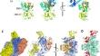
在大肠杆菌和鼠伤寒沙门氏菌中,半胱氨酸的生物合成需要 20 个或更多 cys 基因的产物,这些基因由 CysB 协调调控。在硫限制条件下和诱导剂 N-乙酰丝氨酸存在的情况下,CysB 与 cys 启动子结合,激活下游编码序列的转录。CysB 是一种同源四聚体,由 N 端 DNA 结合结构域(DBD)和 C 端效应结合结构域(EBD)组成。来自产气克雷伯氏菌的 CysB 的二聚体 EBD 片段的晶体结构显示,其蛋白质折叠与 Lac 抑制剂相似,但二聚体的对称性不同,因此 DNA 结合模式并不明显。为了阐明四聚体中的亚基排列,我们测定了完整的 CysB 与 N-乙酰丝氨酸复合体的晶体结构。四聚体中有两种亚基类型,它们的翼螺旋-转螺旋 DNA 结合域与效应结合域的并列位置不同。在组装过程中,四个 EBD 形成核心,DNA 结合域成对排列在表面。N-acetylserine 在一个封闭的结合位点中产生了广泛的极性相互作用,其结合伴随着周围残基的大量构象重排,这些重排传播到蛋白质表面,似乎改变了 DNA 结合域的排列。研究结果(i)与 CysB 现有的大量突变数据进行了讨论,(ii)用于提出 N-乙酰丝氨酸诱导 CysB 激活的结构机制。
The Structure of the LysR-type Transcriptional Regulator, CysB, Bound to the Inducer, N-acetylserine
In Escherichia coli and Salmonella typhimurium, cysteine biosynthesis requires the products of 20 or more cys genes co-ordinately regulated by CysB. Under conditions of sulphur limitation and in the presence of the inducer, N-acetylserine, CysB binds to cys promoters and activates the transcription of the downstream coding sequences. CysB is a homotetramer, comprising an N-terminal DNA binding domain (DBD) and a C-terminal effector binding domain (EBD). The crystal structure of a dimeric EBD fragment of CysB from Klebsiella aerogenes revealed a protein fold similar to that seen in Lac repressor but with a different symmetry in the dimer so that the mode of DNA binding was not apparent. To elucidate the subunit arrangement in the tetramer, we determined the crystal structure of intact CysB in complex with N-acetylserine. The tetramer has two subunit types that differ in the juxtaposition of their winged helix-turn-helix DNA binding domains with respect to the effector binding domain. In the assembly, the four EBDs form a core with the DNA binding domains arranged in pairs on the surface. N-acetylserine makes extensive polar interactions in an enclosed binding site, and its binding is accompanied by substantial conformational rearrangements of surrounding residues that are propagated to the protein surface where they appear to alter the arrangement of the DNA binding domains. The results are (i) discussed in relation to the extensive mutational data available for CysB and (ii) used to propose a structural mechanism of N-acetylserine induced CysB activation.
期刊介绍:
The journal publishes papers in the field of biophysics, which is defined as the study of biological phenomena by using physical methods and concepts. Original papers, reviews and Biophysics letters are published. The primary goal of this journal is to advance the understanding of biological structure and function by application of the principles of physical science, and by presenting the work in a biophysical context.
Papers employing a distinctively biophysical approach at all levels of biological organisation will be considered, as will both experimental and theoretical studies. The criteria for acceptance are scientific content, originality and relevance to biological systems of current interest and importance.
Principal areas of interest include:
- Structure and dynamics of biological macromolecules
- Membrane biophysics and ion channels
- Cell biophysics and organisation
- Macromolecular assemblies
- Biophysical methods and instrumentation
- Advanced microscopics
- System dynamics.
 求助内容:
求助内容: 应助结果提醒方式:


