{"title":"散在肿瘤细胞中的 MUC5AC 免疫反应有助于诊断 CIC 重组肉瘤。","authors":"Shogo Nishino, Naoki Kojima, Hirokazu Sugino, Taisuke Mori, Yasushi Yatabe, Akihiko Yoshida","doi":"10.1007/s00428-024-03863-z","DOIUrl":null,"url":null,"abstract":"<p><p>CIC-rearranged sarcoma is an aggressive round cell sarcoma, and an alternative ATXN1/ATXN1L fusion has been reported. Diagnosis may be difficult, and molecular assays may suffer from imperfect sensitivity. Characteristic histology and ETV4 immunohistochemical positivity are diagnostically helpful. However, ETV4 staining is unavailable in most laboratories. Here, we explored the diagnostic utility of MUC5AC immunohistochemistry in CIC-rearranged sarcomas. All 30 cases, except one, of CIC-rearranged sarcomas and 2 ATXN1-rearranged sarcomas were positive for MUC5AC, although the number of immunopositive cells was generally low (< 5%) in most samples, representing a characteristic scattered pattern. The only MUC5AC-negative case had the lowest tumor volume. Among the 110 mimicking round cell malignancies, 12 tumors showed MUC5AC positivity, including occasional cases of synovial sarcoma and small cell carcinoma, whereas the remaining 98 samples were negative. Despite its lower specificity than that of ETV4 and sparse reactivity that requires careful interpretation, MUC5AC may serve as a useful marker for CIC/ATXN1-rearranged sarcoma because of its wider accessibility.</p>","PeriodicalId":23514,"journal":{"name":"Virchows Archiv","volume":null,"pages":null},"PeriodicalIF":3.4000,"publicationDate":"2024-08-01","publicationTypes":"Journal Article","fieldsOfStudy":null,"isOpenAccess":false,"openAccessPdf":"","citationCount":"0","resultStr":"{\"title\":\"MUC5AC immunoreactivity in scattered tumor cells is useful for diagnosing CIC-rearranged sarcoma.\",\"authors\":\"Shogo Nishino, Naoki Kojima, Hirokazu Sugino, Taisuke Mori, Yasushi Yatabe, Akihiko Yoshida\",\"doi\":\"10.1007/s00428-024-03863-z\",\"DOIUrl\":null,\"url\":null,\"abstract\":\"<p><p>CIC-rearranged sarcoma is an aggressive round cell sarcoma, and an alternative ATXN1/ATXN1L fusion has been reported. Diagnosis may be difficult, and molecular assays may suffer from imperfect sensitivity. Characteristic histology and ETV4 immunohistochemical positivity are diagnostically helpful. However, ETV4 staining is unavailable in most laboratories. Here, we explored the diagnostic utility of MUC5AC immunohistochemistry in CIC-rearranged sarcomas. All 30 cases, except one, of CIC-rearranged sarcomas and 2 ATXN1-rearranged sarcomas were positive for MUC5AC, although the number of immunopositive cells was generally low (< 5%) in most samples, representing a characteristic scattered pattern. The only MUC5AC-negative case had the lowest tumor volume. Among the 110 mimicking round cell malignancies, 12 tumors showed MUC5AC positivity, including occasional cases of synovial sarcoma and small cell carcinoma, whereas the remaining 98 samples were negative. Despite its lower specificity than that of ETV4 and sparse reactivity that requires careful interpretation, MUC5AC may serve as a useful marker for CIC/ATXN1-rearranged sarcoma because of its wider accessibility.</p>\",\"PeriodicalId\":23514,\"journal\":{\"name\":\"Virchows Archiv\",\"volume\":null,\"pages\":null},\"PeriodicalIF\":3.4000,\"publicationDate\":\"2024-08-01\",\"publicationTypes\":\"Journal Article\",\"fieldsOfStudy\":null,\"isOpenAccess\":false,\"openAccessPdf\":\"\",\"citationCount\":\"0\",\"resultStr\":null,\"platform\":\"Semanticscholar\",\"paperid\":null,\"PeriodicalName\":\"Virchows Archiv\",\"FirstCategoryId\":\"3\",\"ListUrlMain\":\"https://doi.org/10.1007/s00428-024-03863-z\",\"RegionNum\":3,\"RegionCategory\":\"医学\",\"ArticlePicture\":[],\"TitleCN\":null,\"AbstractTextCN\":null,\"PMCID\":null,\"EPubDate\":\"2024/7/6 0:00:00\",\"PubModel\":\"Epub\",\"JCR\":\"Q1\",\"JCRName\":\"PATHOLOGY\",\"Score\":null,\"Total\":0}","platform":"Semanticscholar","paperid":null,"PeriodicalName":"Virchows Archiv","FirstCategoryId":"3","ListUrlMain":"https://doi.org/10.1007/s00428-024-03863-z","RegionNum":3,"RegionCategory":"医学","ArticlePicture":[],"TitleCN":null,"AbstractTextCN":null,"PMCID":null,"EPubDate":"2024/7/6 0:00:00","PubModel":"Epub","JCR":"Q1","JCRName":"PATHOLOGY","Score":null,"Total":0}
MUC5AC immunoreactivity in scattered tumor cells is useful for diagnosing CIC-rearranged sarcoma.
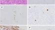
CIC-rearranged sarcoma is an aggressive round cell sarcoma, and an alternative ATXN1/ATXN1L fusion has been reported. Diagnosis may be difficult, and molecular assays may suffer from imperfect sensitivity. Characteristic histology and ETV4 immunohistochemical positivity are diagnostically helpful. However, ETV4 staining is unavailable in most laboratories. Here, we explored the diagnostic utility of MUC5AC immunohistochemistry in CIC-rearranged sarcomas. All 30 cases, except one, of CIC-rearranged sarcomas and 2 ATXN1-rearranged sarcomas were positive for MUC5AC, although the number of immunopositive cells was generally low (< 5%) in most samples, representing a characteristic scattered pattern. The only MUC5AC-negative case had the lowest tumor volume. Among the 110 mimicking round cell malignancies, 12 tumors showed MUC5AC positivity, including occasional cases of synovial sarcoma and small cell carcinoma, whereas the remaining 98 samples were negative. Despite its lower specificity than that of ETV4 and sparse reactivity that requires careful interpretation, MUC5AC may serve as a useful marker for CIC/ATXN1-rearranged sarcoma because of its wider accessibility.
期刊介绍:
Manuscripts of original studies reinforcing the evidence base of modern diagnostic pathology, using immunocytochemical, molecular and ultrastructural techniques, will be welcomed. In addition, papers on critical evaluation of diagnostic criteria but also broadsheets and guidelines with a solid evidence base will be considered. Consideration will also be given to reports of work in other fields relevant to the understanding of human pathology as well as manuscripts on the application of new methods and techniques in pathology. Submission of purely experimental articles is discouraged but manuscripts on experimental work applicable to diagnostic pathology are welcomed. Biomarker studies are welcomed but need to abide by strict rules (e.g. REMARK) of adequate sample size and relevant marker choice. Single marker studies on limited patient series without validated application will as a rule not be considered. Case reports will only be considered when they provide substantial new information with an impact on understanding disease or diagnostic practice.

 求助内容:
求助内容: 应助结果提醒方式:


