Magdalena M. Bolsinger, Alice Drobny, Sibylle Wilfling, Stephanie Reischl, Florian Krach, Raul Moritz, Denise Balta, Ute Hehr, Elisabeth Sock, Florian Bleibaum, Frank Hanses, Beate Winner, Susy Prieto Huarcaya, Philipp Arnold, Friederike Zunke
{"title":"SARS-CoV-2 Spike 蛋白在 HeLa 细胞和肺泡球中诱导时间依赖性 CTSL 上调","authors":"Magdalena M. Bolsinger, Alice Drobny, Sibylle Wilfling, Stephanie Reischl, Florian Krach, Raul Moritz, Denise Balta, Ute Hehr, Elisabeth Sock, Florian Bleibaum, Frank Hanses, Beate Winner, Susy Prieto Huarcaya, Philipp Arnold, Friederike Zunke","doi":"10.1002/jcb.30627","DOIUrl":null,"url":null,"abstract":"<p>Autophagy and lysosomal pathways are involved in the cell entry of SARS-CoV-2 virus. To infect the host cell, the spike protein of SARS-CoV-2 binds to the cell surface receptor angiotensin-converting enzyme 2 (ACE2). To allow the fusion of the viral envelope with the host cell membrane, the spike protein has to be cleaved. One possible mechanism is the endocytosis of the SARS-CoV-2–ACE2 complex and subsequent cleavage of the spike protein, mainly by the lysosomal protease cathepsin L. However, detailed molecular and dynamic insights into the role of cathepsin L in viral cell entry remain elusive. To address this, HeLa cells and iPSC-derived alveolarspheres were treated with recombinant SARS-CoV-2 spike protein, and the changes in mRNA and protein levels of cathepsins L, B, and D were monitored. Additionally, we studied the effect of cathepsin L deficiency on spike protein internalization and investigated the influence of the spike protein on cathepsin L promoters in vitro. Furthermore, we analyzed variants in the genes coding for cathepsin L, B, D, and ACE2 possibly associated with disease progression using data from Regeneron's COVID Results Browser and our own cohort of 173 patients with COVID-19, exhibiting a variant of <i>ACE2</i> showing significant association with COVID-19 disease progression. Our in vitro studies revealed a significant increase in cathepsin L mRNA and protein levels following exposure to the SARS-CoV-2 spike protein in HeLa cells, accompanied by elevated mRNA levels of cathepsin B and D in alveolarspheres. Moreover, an increase in cathepsin L promoter activity was detected in vitro upon spike protein treatment. Notably, the knockout of cathepsin L resulted in reduced internalization of the spike protein. The study highlights the importance of cathepsin L and lysosomal proteases in the SARS-CoV-2 spike protein internalization and suggests the potential of lysosomal proteases as possible therapeutic targets against COVID-19 and other viral infections.</p>","PeriodicalId":15219,"journal":{"name":"Journal of cellular biochemistry","volume":"125 9","pages":""},"PeriodicalIF":3.0000,"publicationDate":"2024-07-07","publicationTypes":"Journal Article","fieldsOfStudy":null,"isOpenAccess":false,"openAccessPdf":"https://onlinelibrary.wiley.com/doi/epdf/10.1002/jcb.30627","citationCount":"0","resultStr":"{\"title\":\"SARS-CoV-2 Spike Protein Induces Time-Dependent CTSL Upregulation in HeLa Cells and Alveolarspheres\",\"authors\":\"Magdalena M. Bolsinger, Alice Drobny, Sibylle Wilfling, Stephanie Reischl, Florian Krach, Raul Moritz, Denise Balta, Ute Hehr, Elisabeth Sock, Florian Bleibaum, Frank Hanses, Beate Winner, Susy Prieto Huarcaya, Philipp Arnold, Friederike Zunke\",\"doi\":\"10.1002/jcb.30627\",\"DOIUrl\":null,\"url\":null,\"abstract\":\"<p>Autophagy and lysosomal pathways are involved in the cell entry of SARS-CoV-2 virus. To infect the host cell, the spike protein of SARS-CoV-2 binds to the cell surface receptor angiotensin-converting enzyme 2 (ACE2). To allow the fusion of the viral envelope with the host cell membrane, the spike protein has to be cleaved. One possible mechanism is the endocytosis of the SARS-CoV-2–ACE2 complex and subsequent cleavage of the spike protein, mainly by the lysosomal protease cathepsin L. However, detailed molecular and dynamic insights into the role of cathepsin L in viral cell entry remain elusive. To address this, HeLa cells and iPSC-derived alveolarspheres were treated with recombinant SARS-CoV-2 spike protein, and the changes in mRNA and protein levels of cathepsins L, B, and D were monitored. Additionally, we studied the effect of cathepsin L deficiency on spike protein internalization and investigated the influence of the spike protein on cathepsin L promoters in vitro. Furthermore, we analyzed variants in the genes coding for cathepsin L, B, D, and ACE2 possibly associated with disease progression using data from Regeneron's COVID Results Browser and our own cohort of 173 patients with COVID-19, exhibiting a variant of <i>ACE2</i> showing significant association with COVID-19 disease progression. Our in vitro studies revealed a significant increase in cathepsin L mRNA and protein levels following exposure to the SARS-CoV-2 spike protein in HeLa cells, accompanied by elevated mRNA levels of cathepsin B and D in alveolarspheres. Moreover, an increase in cathepsin L promoter activity was detected in vitro upon spike protein treatment. Notably, the knockout of cathepsin L resulted in reduced internalization of the spike protein. The study highlights the importance of cathepsin L and lysosomal proteases in the SARS-CoV-2 spike protein internalization and suggests the potential of lysosomal proteases as possible therapeutic targets against COVID-19 and other viral infections.</p>\",\"PeriodicalId\":15219,\"journal\":{\"name\":\"Journal of cellular biochemistry\",\"volume\":\"125 9\",\"pages\":\"\"},\"PeriodicalIF\":3.0000,\"publicationDate\":\"2024-07-07\",\"publicationTypes\":\"Journal Article\",\"fieldsOfStudy\":null,\"isOpenAccess\":false,\"openAccessPdf\":\"https://onlinelibrary.wiley.com/doi/epdf/10.1002/jcb.30627\",\"citationCount\":\"0\",\"resultStr\":null,\"platform\":\"Semanticscholar\",\"paperid\":null,\"PeriodicalName\":\"Journal of cellular biochemistry\",\"FirstCategoryId\":\"99\",\"ListUrlMain\":\"https://onlinelibrary.wiley.com/doi/10.1002/jcb.30627\",\"RegionNum\":3,\"RegionCategory\":\"生物学\",\"ArticlePicture\":[],\"TitleCN\":null,\"AbstractTextCN\":null,\"PMCID\":null,\"EPubDate\":\"\",\"PubModel\":\"\",\"JCR\":\"Q3\",\"JCRName\":\"BIOCHEMISTRY & MOLECULAR BIOLOGY\",\"Score\":null,\"Total\":0}","platform":"Semanticscholar","paperid":null,"PeriodicalName":"Journal of cellular biochemistry","FirstCategoryId":"99","ListUrlMain":"https://onlinelibrary.wiley.com/doi/10.1002/jcb.30627","RegionNum":3,"RegionCategory":"生物学","ArticlePicture":[],"TitleCN":null,"AbstractTextCN":null,"PMCID":null,"EPubDate":"","PubModel":"","JCR":"Q3","JCRName":"BIOCHEMISTRY & MOLECULAR BIOLOGY","Score":null,"Total":0}
引用次数: 0
摘要
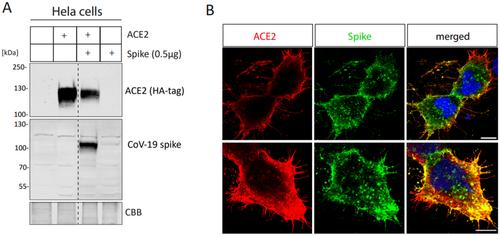
自噬和溶酶体途径参与了 SARS-CoV-2 病毒进入细胞的过程。为了感染宿主细胞,SARS-CoV-2 的尖峰蛋白与细胞表面受体血管紧张素转换酶 2(ACE2)结合。为了使病毒包膜与宿主细胞膜融合,尖峰蛋白必须被裂解。一种可能的机制是,SARS-CoV-2-ACE2 复合物被内吞,随后尖峰蛋白被裂解,主要是被溶酶体蛋白酶 cathepsin L 裂解。为了解决这个问题,我们用重组 SARS-CoV-2 穗状病毒蛋白处理 HeLa 细胞和源自 iPSC 的肺泡球,并监测螯合蛋白 L、B 和 D 的 mRNA 和蛋白水平的变化。此外,我们还研究了螯合蛋白 L 缺乏对尖峰蛋白内化的影响,并在体外研究了尖峰蛋白对螯合蛋白 L 启动子的影响。此外,我们还利用 Regeneron 的 COVID Results Browser 和我们自己的 173 名 COVID-19 患者队列中的数据,分析了可能与疾病进展相关的 cathepsin L、B、D 和 ACE2 编码基因的变异。我们的体外研究显示,在 HeLa 细胞中暴露于 SARS-CoV-2 穗状病毒后,酪蛋白酶 L mRNA 和蛋白水平显著增加,同时肺泡球中酪蛋白酶 B 和 D 的 mRNA 水平也升高。此外,在体外检测到尖峰蛋白处理后, cathepsin L 启动子活性增加。值得注意的是,螯合蛋白 L 的敲除导致尖峰蛋白的内化减少。该研究强调了酪蛋白酶 L 和溶酶体蛋白酶在 SARS-CoV-2 穗状病毒蛋白内化过程中的重要性,并表明溶酶体蛋白酶有可能成为针对 COVID-19 和其他病毒感染的治疗靶点。
SARS-CoV-2 Spike Protein Induces Time-Dependent CTSL Upregulation in HeLa Cells and Alveolarspheres
Autophagy and lysosomal pathways are involved in the cell entry of SARS-CoV-2 virus. To infect the host cell, the spike protein of SARS-CoV-2 binds to the cell surface receptor angiotensin-converting enzyme 2 (ACE2). To allow the fusion of the viral envelope with the host cell membrane, the spike protein has to be cleaved. One possible mechanism is the endocytosis of the SARS-CoV-2–ACE2 complex and subsequent cleavage of the spike protein, mainly by the lysosomal protease cathepsin L. However, detailed molecular and dynamic insights into the role of cathepsin L in viral cell entry remain elusive. To address this, HeLa cells and iPSC-derived alveolarspheres were treated with recombinant SARS-CoV-2 spike protein, and the changes in mRNA and protein levels of cathepsins L, B, and D were monitored. Additionally, we studied the effect of cathepsin L deficiency on spike protein internalization and investigated the influence of the spike protein on cathepsin L promoters in vitro. Furthermore, we analyzed variants in the genes coding for cathepsin L, B, D, and ACE2 possibly associated with disease progression using data from Regeneron's COVID Results Browser and our own cohort of 173 patients with COVID-19, exhibiting a variant of ACE2 showing significant association with COVID-19 disease progression. Our in vitro studies revealed a significant increase in cathepsin L mRNA and protein levels following exposure to the SARS-CoV-2 spike protein in HeLa cells, accompanied by elevated mRNA levels of cathepsin B and D in alveolarspheres. Moreover, an increase in cathepsin L promoter activity was detected in vitro upon spike protein treatment. Notably, the knockout of cathepsin L resulted in reduced internalization of the spike protein. The study highlights the importance of cathepsin L and lysosomal proteases in the SARS-CoV-2 spike protein internalization and suggests the potential of lysosomal proteases as possible therapeutic targets against COVID-19 and other viral infections.
期刊介绍:
The Journal of Cellular Biochemistry publishes descriptions of original research in which complex cellular, pathogenic, clinical, or animal model systems are studied by biochemical, molecular, genetic, epigenetic or quantitative ultrastructural approaches. Submission of papers reporting genomic, proteomic, bioinformatics and systems biology approaches to identify and characterize parameters of biological control in a cellular context are encouraged. The areas covered include, but are not restricted to, conditions, agents, regulatory networks, or differentiation states that influence structure, cell cycle & growth control, structure-function relationships.
 求助内容:
求助内容: 应助结果提醒方式:


