{"title":"上颌骨中心性牙本质鬼细胞瘤:病例报告、新的影像学发现和文献综述。","authors":"Suzuka Yoshida, Yohei Takeshita, Toshiyuki Kawazu, Miki Hisatomi, Shunsuke Okada, Mamiko Fujikura, Kyoichi Obata, Kiyofumi Takabatake, Saori Yoshida, Junichi Asaumi","doi":"10.1007/s11282-024-00764-4","DOIUrl":null,"url":null,"abstract":"<p><p>A dentinogenic ghost cell tumor (DGCT) is a rare benign odontogenic tumor that commonly shows characteristics of solid proliferation and has a relatively high risk of recurrence after surgical treatment. We herein report a case of a central DGCT that occurred in the maxilla and resulted in bone expansion. This study highlights new imaging findings (particularly magnetic resonance imaging) along with histopathological observations. In addition, we conducted a review of the existing literature on this rare tumor. A 37-year-old man developed swelling around the right cheek. A benign odontogenic tumor such as ameloblastoma was suspected based on the imaging examination findings (including bone expansion and the internal characteristics of the tumor) on panoramic imaging, computed tomography, and magnetic resonance imaging. The lesion was surgically excised from the right maxilla. Postoperative histopathological examination led to a definitive diagnosis of central DGCT. The tumor comprised epithelial neoplastic islands, resembling ameloblastoma, inside tight fibroconnective tissue; masses of ghost cells and formation of dentin were also observed. We had suspected that the minute high-density region around the molars on the imaging examinations represented alveolar bone change; however, it represented dentin formation. This led to difficulty diagnosing the lesion. Although DGCT may present characteristic findings on imaging examinations, its occurrence is infrequent, and in some cases, the findings may include the presence or absence of an impacted tooth without obvious calcification. The present case suggests that we should consider the possibility of an odontogenic tumor with calcification when high-density structures are observed inside the lesion.</p>","PeriodicalId":56103,"journal":{"name":"Oral Radiology","volume":" ","pages":"561-568"},"PeriodicalIF":1.6000,"publicationDate":"2024-10-01","publicationTypes":"Journal Article","fieldsOfStudy":null,"isOpenAccess":false,"openAccessPdf":"https://www.ncbi.nlm.nih.gov/pmc/articles/PMC11379793/pdf/","citationCount":"0","resultStr":"{\"title\":\"Central dentinogenic ghost cell tumor of the maxilla: a case report with new imaging findings and review of the literature.\",\"authors\":\"Suzuka Yoshida, Yohei Takeshita, Toshiyuki Kawazu, Miki Hisatomi, Shunsuke Okada, Mamiko Fujikura, Kyoichi Obata, Kiyofumi Takabatake, Saori Yoshida, Junichi Asaumi\",\"doi\":\"10.1007/s11282-024-00764-4\",\"DOIUrl\":null,\"url\":null,\"abstract\":\"<p><p>A dentinogenic ghost cell tumor (DGCT) is a rare benign odontogenic tumor that commonly shows characteristics of solid proliferation and has a relatively high risk of recurrence after surgical treatment. We herein report a case of a central DGCT that occurred in the maxilla and resulted in bone expansion. This study highlights new imaging findings (particularly magnetic resonance imaging) along with histopathological observations. In addition, we conducted a review of the existing literature on this rare tumor. A 37-year-old man developed swelling around the right cheek. A benign odontogenic tumor such as ameloblastoma was suspected based on the imaging examination findings (including bone expansion and the internal characteristics of the tumor) on panoramic imaging, computed tomography, and magnetic resonance imaging. The lesion was surgically excised from the right maxilla. Postoperative histopathological examination led to a definitive diagnosis of central DGCT. The tumor comprised epithelial neoplastic islands, resembling ameloblastoma, inside tight fibroconnective tissue; masses of ghost cells and formation of dentin were also observed. We had suspected that the minute high-density region around the molars on the imaging examinations represented alveolar bone change; however, it represented dentin formation. This led to difficulty diagnosing the lesion. Although DGCT may present characteristic findings on imaging examinations, its occurrence is infrequent, and in some cases, the findings may include the presence or absence of an impacted tooth without obvious calcification. The present case suggests that we should consider the possibility of an odontogenic tumor with calcification when high-density structures are observed inside the lesion.</p>\",\"PeriodicalId\":56103,\"journal\":{\"name\":\"Oral Radiology\",\"volume\":\" \",\"pages\":\"561-568\"},\"PeriodicalIF\":1.6000,\"publicationDate\":\"2024-10-01\",\"publicationTypes\":\"Journal Article\",\"fieldsOfStudy\":null,\"isOpenAccess\":false,\"openAccessPdf\":\"https://www.ncbi.nlm.nih.gov/pmc/articles/PMC11379793/pdf/\",\"citationCount\":\"0\",\"resultStr\":null,\"platform\":\"Semanticscholar\",\"paperid\":null,\"PeriodicalName\":\"Oral Radiology\",\"FirstCategoryId\":\"3\",\"ListUrlMain\":\"https://doi.org/10.1007/s11282-024-00764-4\",\"RegionNum\":3,\"RegionCategory\":\"医学\",\"ArticlePicture\":[],\"TitleCN\":null,\"AbstractTextCN\":null,\"PMCID\":null,\"EPubDate\":\"2024/7/5 0:00:00\",\"PubModel\":\"Epub\",\"JCR\":\"Q3\",\"JCRName\":\"DENTISTRY, ORAL SURGERY & MEDICINE\",\"Score\":null,\"Total\":0}","platform":"Semanticscholar","paperid":null,"PeriodicalName":"Oral Radiology","FirstCategoryId":"3","ListUrlMain":"https://doi.org/10.1007/s11282-024-00764-4","RegionNum":3,"RegionCategory":"医学","ArticlePicture":[],"TitleCN":null,"AbstractTextCN":null,"PMCID":null,"EPubDate":"2024/7/5 0:00:00","PubModel":"Epub","JCR":"Q3","JCRName":"DENTISTRY, ORAL SURGERY & MEDICINE","Score":null,"Total":0}
Central dentinogenic ghost cell tumor of the maxilla: a case report with new imaging findings and review of the literature.
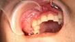
A dentinogenic ghost cell tumor (DGCT) is a rare benign odontogenic tumor that commonly shows characteristics of solid proliferation and has a relatively high risk of recurrence after surgical treatment. We herein report a case of a central DGCT that occurred in the maxilla and resulted in bone expansion. This study highlights new imaging findings (particularly magnetic resonance imaging) along with histopathological observations. In addition, we conducted a review of the existing literature on this rare tumor. A 37-year-old man developed swelling around the right cheek. A benign odontogenic tumor such as ameloblastoma was suspected based on the imaging examination findings (including bone expansion and the internal characteristics of the tumor) on panoramic imaging, computed tomography, and magnetic resonance imaging. The lesion was surgically excised from the right maxilla. Postoperative histopathological examination led to a definitive diagnosis of central DGCT. The tumor comprised epithelial neoplastic islands, resembling ameloblastoma, inside tight fibroconnective tissue; masses of ghost cells and formation of dentin were also observed. We had suspected that the minute high-density region around the molars on the imaging examinations represented alveolar bone change; however, it represented dentin formation. This led to difficulty diagnosing the lesion. Although DGCT may present characteristic findings on imaging examinations, its occurrence is infrequent, and in some cases, the findings may include the presence or absence of an impacted tooth without obvious calcification. The present case suggests that we should consider the possibility of an odontogenic tumor with calcification when high-density structures are observed inside the lesion.
期刊介绍:
As the official English-language journal of the Japanese Society for Oral and Maxillofacial Radiology and the Asian Academy of Oral and Maxillofacial Radiology, Oral Radiology is intended to be a forum for international collaboration in head and neck diagnostic imaging and all related fields. Oral Radiology features cutting-edge research papers, review articles, case reports, and technical notes from both the clinical and experimental fields. As membership in the Society is not a prerequisite, contributions are welcome from researchers and clinicians worldwide.

 求助内容:
求助内容: 应助结果提醒方式:


