Usha D Nagaraj, Jonathan R Dillman, Jean A Tkach, Joshua S Greer, James L Leach
{"title":"利用人工智能图像重建技术评估小儿脑部三维T1加权破坏梯度回波磁共振图像质量。","authors":"Usha D Nagaraj, Jonathan R Dillman, Jean A Tkach, Joshua S Greer, James L Leach","doi":"10.1007/s00234-024-03417-9","DOIUrl":null,"url":null,"abstract":"<p><strong>Purpose: </strong>To assess image quality and diagnostic confidence of 3D T1-weighted spoiled gradient echo (SPGR) MRI using artificial intelligence (AI) reconstruction.</p><p><strong>Materials and methods: </strong>This prospective, IRB-approved study enrolled 50 pediatric patients (mean age = 11.8 ± 3.1 years) undergoing clinical brain MRI. In addition to standard of care (SOC) compressed SENSE (CS = 2.5), 3D T1-weighted SPGR images were obtained with higher CS acceleration factors (5 and 8) to evaluate the ability of AI reconstruction to improve image quality and reduce scan time. Images were reviewed independently on dedicated research PACS workstations by two neuroradiologists. Quantitative analysis of signal intensities to calculate apparent grey and white matter signal to noise (aSNR) and grey-white matter apparent contrast to noise ratios (aCNR) was performed.</p><p><strong>Results: </strong>AI improved overall image quality compared to standard CS reconstruction in 35% (35/100) of evaluations in CS = 2.5 (average scan time = 221 ± 6.9 s), 100% (46/46) of CS = 5 (average scan time = 113.3 ± 4.6 s) and 94% (47/50) of CS = 8 (average scan time = 74.1 ± 0.01 s). Quantitative analysis revealed significantly higher grey matter aSNR, white matter aSNR and grey-white matter aCNR with AI reconstruction compared to standard reconstruction for CS 5 and 8 (all p-values < 0.001), however not for CS 2.5.</p><p><strong>Conclusions: </strong>AI reconstruction improved overall image quality and gray-white matter qualitative and quantitative aSNR and aCNR in highly accelerated (CS = 5 and 8) 3D T1W SPGR images in the majority of pediatric patients.</p>","PeriodicalId":19422,"journal":{"name":"Neuroradiology","volume":" ","pages":"1849-1857"},"PeriodicalIF":2.4000,"publicationDate":"2024-10-01","publicationTypes":"Journal Article","fieldsOfStudy":null,"isOpenAccess":false,"openAccessPdf":"https://www.ncbi.nlm.nih.gov/pmc/articles/PMC11424660/pdf/","citationCount":"0","resultStr":"{\"title\":\"Evaluation of 3D T1-weighted spoiled gradient echo MR image quality using artificial intelligence image reconstruction techniques in the pediatric brain.\",\"authors\":\"Usha D Nagaraj, Jonathan R Dillman, Jean A Tkach, Joshua S Greer, James L Leach\",\"doi\":\"10.1007/s00234-024-03417-9\",\"DOIUrl\":null,\"url\":null,\"abstract\":\"<p><strong>Purpose: </strong>To assess image quality and diagnostic confidence of 3D T1-weighted spoiled gradient echo (SPGR) MRI using artificial intelligence (AI) reconstruction.</p><p><strong>Materials and methods: </strong>This prospective, IRB-approved study enrolled 50 pediatric patients (mean age = 11.8 ± 3.1 years) undergoing clinical brain MRI. In addition to standard of care (SOC) compressed SENSE (CS = 2.5), 3D T1-weighted SPGR images were obtained with higher CS acceleration factors (5 and 8) to evaluate the ability of AI reconstruction to improve image quality and reduce scan time. Images were reviewed independently on dedicated research PACS workstations by two neuroradiologists. Quantitative analysis of signal intensities to calculate apparent grey and white matter signal to noise (aSNR) and grey-white matter apparent contrast to noise ratios (aCNR) was performed.</p><p><strong>Results: </strong>AI improved overall image quality compared to standard CS reconstruction in 35% (35/100) of evaluations in CS = 2.5 (average scan time = 221 ± 6.9 s), 100% (46/46) of CS = 5 (average scan time = 113.3 ± 4.6 s) and 94% (47/50) of CS = 8 (average scan time = 74.1 ± 0.01 s). Quantitative analysis revealed significantly higher grey matter aSNR, white matter aSNR and grey-white matter aCNR with AI reconstruction compared to standard reconstruction for CS 5 and 8 (all p-values < 0.001), however not for CS 2.5.</p><p><strong>Conclusions: </strong>AI reconstruction improved overall image quality and gray-white matter qualitative and quantitative aSNR and aCNR in highly accelerated (CS = 5 and 8) 3D T1W SPGR images in the majority of pediatric patients.</p>\",\"PeriodicalId\":19422,\"journal\":{\"name\":\"Neuroradiology\",\"volume\":\" \",\"pages\":\"1849-1857\"},\"PeriodicalIF\":2.4000,\"publicationDate\":\"2024-10-01\",\"publicationTypes\":\"Journal Article\",\"fieldsOfStudy\":null,\"isOpenAccess\":false,\"openAccessPdf\":\"https://www.ncbi.nlm.nih.gov/pmc/articles/PMC11424660/pdf/\",\"citationCount\":\"0\",\"resultStr\":null,\"platform\":\"Semanticscholar\",\"paperid\":null,\"PeriodicalName\":\"Neuroradiology\",\"FirstCategoryId\":\"3\",\"ListUrlMain\":\"https://doi.org/10.1007/s00234-024-03417-9\",\"RegionNum\":3,\"RegionCategory\":\"医学\",\"ArticlePicture\":[],\"TitleCN\":null,\"AbstractTextCN\":null,\"PMCID\":null,\"EPubDate\":\"2024/7/5 0:00:00\",\"PubModel\":\"Epub\",\"JCR\":\"Q2\",\"JCRName\":\"CLINICAL NEUROLOGY\",\"Score\":null,\"Total\":0}","platform":"Semanticscholar","paperid":null,"PeriodicalName":"Neuroradiology","FirstCategoryId":"3","ListUrlMain":"https://doi.org/10.1007/s00234-024-03417-9","RegionNum":3,"RegionCategory":"医学","ArticlePicture":[],"TitleCN":null,"AbstractTextCN":null,"PMCID":null,"EPubDate":"2024/7/5 0:00:00","PubModel":"Epub","JCR":"Q2","JCRName":"CLINICAL NEUROLOGY","Score":null,"Total":0}
Evaluation of 3D T1-weighted spoiled gradient echo MR image quality using artificial intelligence image reconstruction techniques in the pediatric brain.
Purpose: To assess image quality and diagnostic confidence of 3D T1-weighted spoiled gradient echo (SPGR) MRI using artificial intelligence (AI) reconstruction.
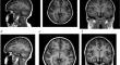
Materials and methods: This prospective, IRB-approved study enrolled 50 pediatric patients (mean age = 11.8 ± 3.1 years) undergoing clinical brain MRI. In addition to standard of care (SOC) compressed SENSE (CS = 2.5), 3D T1-weighted SPGR images were obtained with higher CS acceleration factors (5 and 8) to evaluate the ability of AI reconstruction to improve image quality and reduce scan time. Images were reviewed independently on dedicated research PACS workstations by two neuroradiologists. Quantitative analysis of signal intensities to calculate apparent grey and white matter signal to noise (aSNR) and grey-white matter apparent contrast to noise ratios (aCNR) was performed.
Results: AI improved overall image quality compared to standard CS reconstruction in 35% (35/100) of evaluations in CS = 2.5 (average scan time = 221 ± 6.9 s), 100% (46/46) of CS = 5 (average scan time = 113.3 ± 4.6 s) and 94% (47/50) of CS = 8 (average scan time = 74.1 ± 0.01 s). Quantitative analysis revealed significantly higher grey matter aSNR, white matter aSNR and grey-white matter aCNR with AI reconstruction compared to standard reconstruction for CS 5 and 8 (all p-values < 0.001), however not for CS 2.5.
Conclusions: AI reconstruction improved overall image quality and gray-white matter qualitative and quantitative aSNR and aCNR in highly accelerated (CS = 5 and 8) 3D T1W SPGR images in the majority of pediatric patients.
期刊介绍:
Neuroradiology aims to provide state-of-the-art medical and scientific information in the fields of Neuroradiology, Neurosciences, Neurology, Psychiatry, Neurosurgery, and related medical specialities. Neuroradiology as the official Journal of the European Society of Neuroradiology receives submissions from all parts of the world and publishes peer-reviewed original research, comprehensive reviews, educational papers, opinion papers, and short reports on exceptional clinical observations and new technical developments in the field of Neuroimaging and Neurointervention. The journal has subsections for Diagnostic and Interventional Neuroradiology, Advanced Neuroimaging, Paediatric Neuroradiology, Head-Neck-ENT Radiology, Spine Neuroradiology, and for submissions from Japan. Neuroradiology aims to provide new knowledge about and insights into the function and pathology of the human nervous system that may help to better diagnose and treat nervous system diseases. Neuroradiology is a member of the Committee on Publication Ethics (COPE) and follows the COPE core practices. Neuroradiology prefers articles that are free of bias, self-critical regarding limitations, transparent and clear in describing study participants, methods, and statistics, and short in presenting results. Before peer-review all submissions are automatically checked by iThenticate to assess for potential overlap in prior publication.

 求助内容:
求助内容: 应助结果提醒方式:


