A. A. Veshchitskii, A. V. Belyaev, N. S. Merkulyeva
{"title":"新生猫和成年猫脊髓中副发光素-免疫阳性神经元的分布比较","authors":"A. A. Veshchitskii, A. V. Belyaev, N. S. Merkulyeva","doi":"10.1134/s0022093024030049","DOIUrl":null,"url":null,"abstract":"<h3 data-test=\"abstract-sub-heading\">Abstract</h3><p>We performed a comparative immunohistochemical analysis of\nparvalbumin, a calcium-binding protein, expression in the lumbosacral\nspinal cord of newborn and adult cats. In contrast to adult animals,\nparvalbumin immunostaining in newborn kittens was mainly detected\nin the sensory fibers located in the dorsal horns and medial part\nof the intermediate gray matter. The location of these fibers partially\nrecapitulates the position of the Clarke’s nuclei, but is not limited\nby their classical boundaries, spanning the entire length of the\nlumbar spinal cord and transitioning into the putative Stilling’s\nnuclei in the sacral region. Thus, in newborn kittens, in contrast\nto adult animals, parvalbumin-immunopositive proprioceptive fibers\nappear to make up a single network. We hypothesize that with age,\nthis continuous system of fibers undergoes restructuring due to\nmaturation and spreading of the neuronal and neuropil components\nof the lumbar enlargement, responsible primarily for the locomotor\ncontrol. As a result, two local groups of interneurons, the Clarke’s\nand Stilling’s nuclei, form. The only parvalbumin-immunopositive\nspinal neurons in newborn kittens are premotor interneurons located\nalong the lamina IX curvature. These neurons are characterized by\na weak or no immunolabeling of the neuronal marker protein NeuN,\nwhich is indicative of a special neurochemical status of these neurons.</p>","PeriodicalId":15805,"journal":{"name":"Journal of Evolutionary Biochemistry and Physiology","volume":"31 1","pages":""},"PeriodicalIF":0.5000,"publicationDate":"2024-06-26","publicationTypes":"Journal Article","fieldsOfStudy":null,"isOpenAccess":false,"openAccessPdf":"","citationCount":"0","resultStr":"{\"title\":\"A Comparative Distribution of Parvalbumin-Immunopositive Neural Elements in the Spinal Cord of Newborn and Adult Cats\",\"authors\":\"A. A. Veshchitskii, A. V. Belyaev, N. S. Merkulyeva\",\"doi\":\"10.1134/s0022093024030049\",\"DOIUrl\":null,\"url\":null,\"abstract\":\"<h3 data-test=\\\"abstract-sub-heading\\\">Abstract</h3><p>We performed a comparative immunohistochemical analysis of\\nparvalbumin, a calcium-binding protein, expression in the lumbosacral\\nspinal cord of newborn and adult cats. In contrast to adult animals,\\nparvalbumin immunostaining in newborn kittens was mainly detected\\nin the sensory fibers located in the dorsal horns and medial part\\nof the intermediate gray matter. The location of these fibers partially\\nrecapitulates the position of the Clarke’s nuclei, but is not limited\\nby their classical boundaries, spanning the entire length of the\\nlumbar spinal cord and transitioning into the putative Stilling’s\\nnuclei in the sacral region. Thus, in newborn kittens, in contrast\\nto adult animals, parvalbumin-immunopositive proprioceptive fibers\\nappear to make up a single network. We hypothesize that with age,\\nthis continuous system of fibers undergoes restructuring due to\\nmaturation and spreading of the neuronal and neuropil components\\nof the lumbar enlargement, responsible primarily for the locomotor\\ncontrol. As a result, two local groups of interneurons, the Clarke’s\\nand Stilling’s nuclei, form. The only parvalbumin-immunopositive\\nspinal neurons in newborn kittens are premotor interneurons located\\nalong the lamina IX curvature. These neurons are characterized by\\na weak or no immunolabeling of the neuronal marker protein NeuN,\\nwhich is indicative of a special neurochemical status of these neurons.</p>\",\"PeriodicalId\":15805,\"journal\":{\"name\":\"Journal of Evolutionary Biochemistry and Physiology\",\"volume\":\"31 1\",\"pages\":\"\"},\"PeriodicalIF\":0.5000,\"publicationDate\":\"2024-06-26\",\"publicationTypes\":\"Journal Article\",\"fieldsOfStudy\":null,\"isOpenAccess\":false,\"openAccessPdf\":\"\",\"citationCount\":\"0\",\"resultStr\":null,\"platform\":\"Semanticscholar\",\"paperid\":null,\"PeriodicalName\":\"Journal of Evolutionary Biochemistry and Physiology\",\"FirstCategoryId\":\"99\",\"ListUrlMain\":\"https://doi.org/10.1134/s0022093024030049\",\"RegionNum\":4,\"RegionCategory\":\"生物学\",\"ArticlePicture\":[],\"TitleCN\":null,\"AbstractTextCN\":null,\"PMCID\":null,\"EPubDate\":\"\",\"PubModel\":\"\",\"JCR\":\"Q4\",\"JCRName\":\"BIOCHEMISTRY & MOLECULAR BIOLOGY\",\"Score\":null,\"Total\":0}","platform":"Semanticscholar","paperid":null,"PeriodicalName":"Journal of Evolutionary Biochemistry and Physiology","FirstCategoryId":"99","ListUrlMain":"https://doi.org/10.1134/s0022093024030049","RegionNum":4,"RegionCategory":"生物学","ArticlePicture":[],"TitleCN":null,"AbstractTextCN":null,"PMCID":null,"EPubDate":"","PubModel":"","JCR":"Q4","JCRName":"BIOCHEMISTRY & MOLECULAR BIOLOGY","Score":null,"Total":0}
A Comparative Distribution of Parvalbumin-Immunopositive Neural Elements in the Spinal Cord of Newborn and Adult Cats
Abstract
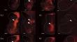
We performed a comparative immunohistochemical analysis of
parvalbumin, a calcium-binding protein, expression in the lumbosacral
spinal cord of newborn and adult cats. In contrast to adult animals,
parvalbumin immunostaining in newborn kittens was mainly detected
in the sensory fibers located in the dorsal horns and medial part
of the intermediate gray matter. The location of these fibers partially
recapitulates the position of the Clarke’s nuclei, but is not limited
by their classical boundaries, spanning the entire length of the
lumbar spinal cord and transitioning into the putative Stilling’s
nuclei in the sacral region. Thus, in newborn kittens, in contrast
to adult animals, parvalbumin-immunopositive proprioceptive fibers
appear to make up a single network. We hypothesize that with age,
this continuous system of fibers undergoes restructuring due to
maturation and spreading of the neuronal and neuropil components
of the lumbar enlargement, responsible primarily for the locomotor
control. As a result, two local groups of interneurons, the Clarke’s
and Stilling’s nuclei, form. The only parvalbumin-immunopositive
spinal neurons in newborn kittens are premotor interneurons located
along the lamina IX curvature. These neurons are characterized by
a weak or no immunolabeling of the neuronal marker protein NeuN,
which is indicative of a special neurochemical status of these neurons.
期刊介绍:
Journal of Evolutionary Biochemistry and Physiology publishes original experimental and theoretical and review articles related to evolution of the main forms of metabolism in connection with life origin; comparative and ontogenetic physiology and biochemistry, biochemical evolution of animal world; as well as evolution of functions; morphology, pharmacology, pathophysiology and ecological physiology. The journal welcomes manuscripts from all countries in the English or Russian language.
 求助内容:
求助内容: 应助结果提醒方式:


