J. D. Korobkina, M. A. Panteleev, A. N. Sveshnikova
{"title":"人类和鼠类精子钙反应机制的比较研究","authors":"J. D. Korobkina, M. A. Panteleev, A. N. Sveshnikova","doi":"10.1134/S1990747824700132","DOIUrl":null,"url":null,"abstract":"<p>Calcium signaling is a principal method of signal transduction in cells of non-excitable tissues. In both mouse and human sperm, it can be induced in response to progesterone, manifesting as oscillations or single peaks and followed by the acrosomal reaction. However, the molecular mechanisms of progesterone activation may vary between species. In this study, we aim to compare the calcium signaling mechanisms in human and mouse spermatozoa. We investigated the calcium response in mouse sperm activated by progesterone. We employed spectrofluorometry to quantify the rise in calcium concentration in response to progesterone in Fura-2 loaded mouse sperm cells in suspension. Our experiments demonstrated that mouse sperm cells respond to 50 μM progesterone with a peak 120 ± 35 s wide and 0.8 ± 0.3 μM high. Based on literature data, a scheme for the induction of calcium signaling was constructed, suggesting an intermediate stage with the synthesis of a certain prostanoid (possibly PGE2) and activation of mouse sperm by this prostanoid through a G-protein-coupled receptor. Based on the obtained reaction scheme, two computational models were developed: a point model and a three-dimensional model. As with human sperm, the point model provided only a qualitative description of calcium responses, whereas the three-dimensional model produced the shape of the calcium peak and the frequency of calcium oscillations in response to progesterone that were similar to the experimentally obtained values. Using in silico analysis, it was shown that in mouse sperm, the spatial distribution of signaling enzymes regulates the type and form of the calcium response. We conclude that the presence of time delays due to the diffusion and spatial distribution of calcium signaling enzymes regulates the calcium response in both human and mouse sperm.</p>","PeriodicalId":484,"journal":{"name":"Biochemistry (Moscow), Supplement Series A: Membrane and Cell Biology","volume":"18 2","pages":"110 - 126"},"PeriodicalIF":1.1000,"publicationDate":"2024-06-25","publicationTypes":"Journal Article","fieldsOfStudy":null,"isOpenAccess":false,"openAccessPdf":"","citationCount":"0","resultStr":"{\"title\":\"Comparative Investigation of the Mechanisms of Calcium Response in Human and Murine Spermatozoa\",\"authors\":\"J. D. Korobkina, M. A. Panteleev, A. N. Sveshnikova\",\"doi\":\"10.1134/S1990747824700132\",\"DOIUrl\":null,\"url\":null,\"abstract\":\"<p>Calcium signaling is a principal method of signal transduction in cells of non-excitable tissues. In both mouse and human sperm, it can be induced in response to progesterone, manifesting as oscillations or single peaks and followed by the acrosomal reaction. However, the molecular mechanisms of progesterone activation may vary between species. In this study, we aim to compare the calcium signaling mechanisms in human and mouse spermatozoa. We investigated the calcium response in mouse sperm activated by progesterone. We employed spectrofluorometry to quantify the rise in calcium concentration in response to progesterone in Fura-2 loaded mouse sperm cells in suspension. Our experiments demonstrated that mouse sperm cells respond to 50 μM progesterone with a peak 120 ± 35 s wide and 0.8 ± 0.3 μM high. Based on literature data, a scheme for the induction of calcium signaling was constructed, suggesting an intermediate stage with the synthesis of a certain prostanoid (possibly PGE2) and activation of mouse sperm by this prostanoid through a G-protein-coupled receptor. Based on the obtained reaction scheme, two computational models were developed: a point model and a three-dimensional model. As with human sperm, the point model provided only a qualitative description of calcium responses, whereas the three-dimensional model produced the shape of the calcium peak and the frequency of calcium oscillations in response to progesterone that were similar to the experimentally obtained values. Using in silico analysis, it was shown that in mouse sperm, the spatial distribution of signaling enzymes regulates the type and form of the calcium response. We conclude that the presence of time delays due to the diffusion and spatial distribution of calcium signaling enzymes regulates the calcium response in both human and mouse sperm.</p>\",\"PeriodicalId\":484,\"journal\":{\"name\":\"Biochemistry (Moscow), Supplement Series A: Membrane and Cell Biology\",\"volume\":\"18 2\",\"pages\":\"110 - 126\"},\"PeriodicalIF\":1.1000,\"publicationDate\":\"2024-06-25\",\"publicationTypes\":\"Journal Article\",\"fieldsOfStudy\":null,\"isOpenAccess\":false,\"openAccessPdf\":\"\",\"citationCount\":\"0\",\"resultStr\":null,\"platform\":\"Semanticscholar\",\"paperid\":null,\"PeriodicalName\":\"Biochemistry (Moscow), Supplement Series A: Membrane and Cell Biology\",\"FirstCategoryId\":\"2\",\"ListUrlMain\":\"https://link.springer.com/article/10.1134/S1990747824700132\",\"RegionNum\":0,\"RegionCategory\":null,\"ArticlePicture\":[],\"TitleCN\":null,\"AbstractTextCN\":null,\"PMCID\":null,\"EPubDate\":\"\",\"PubModel\":\"\",\"JCR\":\"Q4\",\"JCRName\":\"CELL BIOLOGY\",\"Score\":null,\"Total\":0}","platform":"Semanticscholar","paperid":null,"PeriodicalName":"Biochemistry (Moscow), Supplement Series A: Membrane and Cell Biology","FirstCategoryId":"2","ListUrlMain":"https://link.springer.com/article/10.1134/S1990747824700132","RegionNum":0,"RegionCategory":null,"ArticlePicture":[],"TitleCN":null,"AbstractTextCN":null,"PMCID":null,"EPubDate":"","PubModel":"","JCR":"Q4","JCRName":"CELL BIOLOGY","Score":null,"Total":0}
Comparative Investigation of the Mechanisms of Calcium Response in Human and Murine Spermatozoa
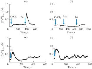
Calcium signaling is a principal method of signal transduction in cells of non-excitable tissues. In both mouse and human sperm, it can be induced in response to progesterone, manifesting as oscillations or single peaks and followed by the acrosomal reaction. However, the molecular mechanisms of progesterone activation may vary between species. In this study, we aim to compare the calcium signaling mechanisms in human and mouse spermatozoa. We investigated the calcium response in mouse sperm activated by progesterone. We employed spectrofluorometry to quantify the rise in calcium concentration in response to progesterone in Fura-2 loaded mouse sperm cells in suspension. Our experiments demonstrated that mouse sperm cells respond to 50 μM progesterone with a peak 120 ± 35 s wide and 0.8 ± 0.3 μM high. Based on literature data, a scheme for the induction of calcium signaling was constructed, suggesting an intermediate stage with the synthesis of a certain prostanoid (possibly PGE2) and activation of mouse sperm by this prostanoid through a G-protein-coupled receptor. Based on the obtained reaction scheme, two computational models were developed: a point model and a three-dimensional model. As with human sperm, the point model provided only a qualitative description of calcium responses, whereas the three-dimensional model produced the shape of the calcium peak and the frequency of calcium oscillations in response to progesterone that were similar to the experimentally obtained values. Using in silico analysis, it was shown that in mouse sperm, the spatial distribution of signaling enzymes regulates the type and form of the calcium response. We conclude that the presence of time delays due to the diffusion and spatial distribution of calcium signaling enzymes regulates the calcium response in both human and mouse sperm.
期刊介绍:
Biochemistry (Moscow), Supplement Series A: Membrane and Cell Biology is an international peer reviewed journal that publishes original articles on physical, chemical, and molecular mechanisms that underlie basic properties of biological membranes and mediate membrane-related cellular functions. The primary topics of the journal are membrane structure, mechanisms of membrane transport, bioenergetics and photobiology, intracellular signaling as well as membrane aspects of cell biology, immunology, and medicine. The journal is multidisciplinary and gives preference to those articles that employ a variety of experimental approaches, basically in biophysics but also in biochemistry, cytology, and molecular biology. The journal publishes articles that strive for unveiling membrane and cellular functions through innovative theoretical models and computer simulations.
 求助内容:
求助内容: 应助结果提醒方式:


