Mohammad Jafar Bagheri, Mojtaba Rezazadeh Valojerdi, Mojdeh Salehnia
{"title":"人子宫内膜间充质干细胞与小鼠卵母细胞在三维培养系统中共同培养形成卵巢类器官","authors":"Mohammad Jafar Bagheri, Mojtaba Rezazadeh Valojerdi, Mojdeh Salehnia","doi":"10.1007/s10616-024-00639-w","DOIUrl":null,"url":null,"abstract":"<p>The purpose of this study was to compare the formation of organoid structures by co-culturing of human endometrial mesenchymal stem cells (hEnMSCs) and mouse germinal vesicle (GV) oocytes in hanging drop and sodium alginate hydrogel co-culture methods. Following the preparation of hEnMSCs and partially denuded mouse germinal vesicle oocytes, they were co-cultured in hanging drop and sodium alginate hydrogel systems as two experimental groups. In respected control groups the hEnMSCs were cultured without oocytes. The organoid formation was evaluated under the inverted microscope in all studied groups during the culture period. The hematoxylin and eosin, alcian blue, periodic acid Schiff, and Masson's trichrome methods, were applied for morphological evaluation and extracellular matrix components staining such as glycosaminoglycan, carbohydrate, and collagen fibers. In addition, the germ cell-like characteristics within the organoid structures were investigated via alkaline phosphatase activity immunocytochemistry for DEAD-box polypeptide 4 (DDX4), and the expression of octamer-binding transcription factor 4 (OCT4), DDX4, and synaptonemal complex protein 3 (SYCP3) genes by real-time RT-PCR. The culturing of hEnMSCs in the hanging drop method led to the formation of organoid structures while this structure was not seen in sodium alginate hydrogel culture. The mean diameter of organoid structures was increased during 4 days of culture in both the experimental and control groups in the hanging drop method, reaching 675.50 ± 18.55 µm and 670.25 ± 21.40 µm, respectively (P < 0.05). Morphological staining indicated some large ovoid cells with euchromatin nuclei in the experimental group, whereas, in the control group cells showed dark and dense nuclei. The extracellular matrix components were deposited in organoid structures in both control and experimental groups. The positive alkaline phosphatase activity and immunocytochemistry for DDX4 confirmed the presence of germ cell-like in the experimental group. Real-time RT-PCR showed a significant increase in the expression of DDX4 and SYCP3 genes and a decrease in the level of OCT4 expression in the experimental group compared with its controls. This study successfully generated organoid structures by co-culture of hEnMSCs and oocytes in the hanging drop method and the hEnMSCs could be differentiated into germ cell-like. This organoid structure has potential applications in regenerative medicine and reproductive biology.</p>","PeriodicalId":10890,"journal":{"name":"Cytotechnology","volume":"174 1","pages":""},"PeriodicalIF":2.0000,"publicationDate":"2024-06-24","publicationTypes":"Journal Article","fieldsOfStudy":null,"isOpenAccess":false,"openAccessPdf":"","citationCount":"0","resultStr":"{\"title\":\"Formation of ovarian organoid by co-culture of human endometrial mesenchymal stem cells and mouse oocyte in 3-dimensional culture system\",\"authors\":\"Mohammad Jafar Bagheri, Mojtaba Rezazadeh Valojerdi, Mojdeh Salehnia\",\"doi\":\"10.1007/s10616-024-00639-w\",\"DOIUrl\":null,\"url\":null,\"abstract\":\"<p>The purpose of this study was to compare the formation of organoid structures by co-culturing of human endometrial mesenchymal stem cells (hEnMSCs) and mouse germinal vesicle (GV) oocytes in hanging drop and sodium alginate hydrogel co-culture methods. Following the preparation of hEnMSCs and partially denuded mouse germinal vesicle oocytes, they were co-cultured in hanging drop and sodium alginate hydrogel systems as two experimental groups. In respected control groups the hEnMSCs were cultured without oocytes. The organoid formation was evaluated under the inverted microscope in all studied groups during the culture period. The hematoxylin and eosin, alcian blue, periodic acid Schiff, and Masson's trichrome methods, were applied for morphological evaluation and extracellular matrix components staining such as glycosaminoglycan, carbohydrate, and collagen fibers. In addition, the germ cell-like characteristics within the organoid structures were investigated via alkaline phosphatase activity immunocytochemistry for DEAD-box polypeptide 4 (DDX4), and the expression of octamer-binding transcription factor 4 (OCT4), DDX4, and synaptonemal complex protein 3 (SYCP3) genes by real-time RT-PCR. The culturing of hEnMSCs in the hanging drop method led to the formation of organoid structures while this structure was not seen in sodium alginate hydrogel culture. The mean diameter of organoid structures was increased during 4 days of culture in both the experimental and control groups in the hanging drop method, reaching 675.50 ± 18.55 µm and 670.25 ± 21.40 µm, respectively (P < 0.05). Morphological staining indicated some large ovoid cells with euchromatin nuclei in the experimental group, whereas, in the control group cells showed dark and dense nuclei. The extracellular matrix components were deposited in organoid structures in both control and experimental groups. The positive alkaline phosphatase activity and immunocytochemistry for DDX4 confirmed the presence of germ cell-like in the experimental group. Real-time RT-PCR showed a significant increase in the expression of DDX4 and SYCP3 genes and a decrease in the level of OCT4 expression in the experimental group compared with its controls. This study successfully generated organoid structures by co-culture of hEnMSCs and oocytes in the hanging drop method and the hEnMSCs could be differentiated into germ cell-like. This organoid structure has potential applications in regenerative medicine and reproductive biology.</p>\",\"PeriodicalId\":10890,\"journal\":{\"name\":\"Cytotechnology\",\"volume\":\"174 1\",\"pages\":\"\"},\"PeriodicalIF\":2.0000,\"publicationDate\":\"2024-06-24\",\"publicationTypes\":\"Journal Article\",\"fieldsOfStudy\":null,\"isOpenAccess\":false,\"openAccessPdf\":\"\",\"citationCount\":\"0\",\"resultStr\":null,\"platform\":\"Semanticscholar\",\"paperid\":null,\"PeriodicalName\":\"Cytotechnology\",\"FirstCategoryId\":\"99\",\"ListUrlMain\":\"https://doi.org/10.1007/s10616-024-00639-w\",\"RegionNum\":4,\"RegionCategory\":\"生物学\",\"ArticlePicture\":[],\"TitleCN\":null,\"AbstractTextCN\":null,\"PMCID\":null,\"EPubDate\":\"\",\"PubModel\":\"\",\"JCR\":\"Q3\",\"JCRName\":\"BIOTECHNOLOGY & APPLIED MICROBIOLOGY\",\"Score\":null,\"Total\":0}","platform":"Semanticscholar","paperid":null,"PeriodicalName":"Cytotechnology","FirstCategoryId":"99","ListUrlMain":"https://doi.org/10.1007/s10616-024-00639-w","RegionNum":4,"RegionCategory":"生物学","ArticlePicture":[],"TitleCN":null,"AbstractTextCN":null,"PMCID":null,"EPubDate":"","PubModel":"","JCR":"Q3","JCRName":"BIOTECHNOLOGY & APPLIED MICROBIOLOGY","Score":null,"Total":0}
Formation of ovarian organoid by co-culture of human endometrial mesenchymal stem cells and mouse oocyte in 3-dimensional culture system
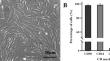
The purpose of this study was to compare the formation of organoid structures by co-culturing of human endometrial mesenchymal stem cells (hEnMSCs) and mouse germinal vesicle (GV) oocytes in hanging drop and sodium alginate hydrogel co-culture methods. Following the preparation of hEnMSCs and partially denuded mouse germinal vesicle oocytes, they were co-cultured in hanging drop and sodium alginate hydrogel systems as two experimental groups. In respected control groups the hEnMSCs were cultured without oocytes. The organoid formation was evaluated under the inverted microscope in all studied groups during the culture period. The hematoxylin and eosin, alcian blue, periodic acid Schiff, and Masson's trichrome methods, were applied for morphological evaluation and extracellular matrix components staining such as glycosaminoglycan, carbohydrate, and collagen fibers. In addition, the germ cell-like characteristics within the organoid structures were investigated via alkaline phosphatase activity immunocytochemistry for DEAD-box polypeptide 4 (DDX4), and the expression of octamer-binding transcription factor 4 (OCT4), DDX4, and synaptonemal complex protein 3 (SYCP3) genes by real-time RT-PCR. The culturing of hEnMSCs in the hanging drop method led to the formation of organoid structures while this structure was not seen in sodium alginate hydrogel culture. The mean diameter of organoid structures was increased during 4 days of culture in both the experimental and control groups in the hanging drop method, reaching 675.50 ± 18.55 µm and 670.25 ± 21.40 µm, respectively (P < 0.05). Morphological staining indicated some large ovoid cells with euchromatin nuclei in the experimental group, whereas, in the control group cells showed dark and dense nuclei. The extracellular matrix components were deposited in organoid structures in both control and experimental groups. The positive alkaline phosphatase activity and immunocytochemistry for DDX4 confirmed the presence of germ cell-like in the experimental group. Real-time RT-PCR showed a significant increase in the expression of DDX4 and SYCP3 genes and a decrease in the level of OCT4 expression in the experimental group compared with its controls. This study successfully generated organoid structures by co-culture of hEnMSCs and oocytes in the hanging drop method and the hEnMSCs could be differentiated into germ cell-like. This organoid structure has potential applications in regenerative medicine and reproductive biology.
期刊介绍:
The scope of the Journal includes:
1. The derivation, genetic modification and characterization of cell lines, genetic and phenotypic regulation, control of cellular metabolism, cell physiology and biochemistry related to cell function, performance and expression of cell products.
2. Cell culture techniques, substrates, environmental requirements and optimization, cloning, hybridization and molecular biology, including genomic and proteomic tools.
3. Cell culture systems, processes, reactors, scale-up, and industrial production. Descriptions of the design or construction of equipment, media or quality control procedures, that are ancillary to cellular research.
4. The application of animal/human cells in research in the field of stem cell research including maintenance of stemness, differentiation, genetics, and senescence, cancer research, research in immunology, as well as applications in tissue engineering and gene therapy.
5. The use of cell cultures as a substrate for bioassays, biomedical applications and in particular as a replacement for animal models.
 求助内容:
求助内容: 应助结果提醒方式:


