Sandryne David, Nassim Ksantini, Frédérick Dallaire, Katherine Ember, François Daoust, Guillaume Sheehy, Costas G. Hadjipanayis, Kevin Petrecca, Brian C. Wilson, Frédéric Leblond
{"title":"利用多光谱非弹性散射检测技术实现多发性癌症的非接触式宏观成像。","authors":"Sandryne David, Nassim Ksantini, Frédérick Dallaire, Katherine Ember, François Daoust, Guillaume Sheehy, Costas G. Hadjipanayis, Kevin Petrecca, Brian C. Wilson, Frédéric Leblond","doi":"10.1002/jbio.202400087","DOIUrl":null,"url":null,"abstract":"<p>Here we introduce a Raman spectroscopy approach combining multi-spectral imaging and a new fluorescence background subtraction technique to image individual Raman peaks in less than 5 seconds over a square field-of-view of 1-centimeter sides with 350 micrometers resolution. First, human data is presented supporting the feasibility of achieving cancer detection with high sensitivity and specificity – in brain, breast, lung, and ovarian/endometrium tissue – using no more than three biochemically interpretable biomarkers associated with the inelastic scattering signal from specific Raman peaks. Second, a proof-of-principle study in biological tissue is presented demonstrating the feasibility of detecting a single Raman band – here the CH<sub>2</sub>/CH<sub>3</sub> deformation bands from proteins and lipids – using a conventional multi-spectral imaging system in combination with the new background removal method. This study paves the way for the development of a new Raman imaging technique that is rapid, label-free, and wide field.</p>","PeriodicalId":184,"journal":{"name":"Journal of Biophotonics","volume":"17 9","pages":""},"PeriodicalIF":2.0000,"publicationDate":"2024-07-04","publicationTypes":"Journal Article","fieldsOfStudy":null,"isOpenAccess":false,"openAccessPdf":"https://onlinelibrary.wiley.com/doi/epdf/10.1002/jbio.202400087","citationCount":"0","resultStr":"{\"title\":\"Toward noncontact macroscopic imaging of multiple cancers using multi-spectral inelastic scattering detection\",\"authors\":\"Sandryne David, Nassim Ksantini, Frédérick Dallaire, Katherine Ember, François Daoust, Guillaume Sheehy, Costas G. Hadjipanayis, Kevin Petrecca, Brian C. Wilson, Frédéric Leblond\",\"doi\":\"10.1002/jbio.202400087\",\"DOIUrl\":null,\"url\":null,\"abstract\":\"<p>Here we introduce a Raman spectroscopy approach combining multi-spectral imaging and a new fluorescence background subtraction technique to image individual Raman peaks in less than 5 seconds over a square field-of-view of 1-centimeter sides with 350 micrometers resolution. First, human data is presented supporting the feasibility of achieving cancer detection with high sensitivity and specificity – in brain, breast, lung, and ovarian/endometrium tissue – using no more than three biochemically interpretable biomarkers associated with the inelastic scattering signal from specific Raman peaks. Second, a proof-of-principle study in biological tissue is presented demonstrating the feasibility of detecting a single Raman band – here the CH<sub>2</sub>/CH<sub>3</sub> deformation bands from proteins and lipids – using a conventional multi-spectral imaging system in combination with the new background removal method. This study paves the way for the development of a new Raman imaging technique that is rapid, label-free, and wide field.</p>\",\"PeriodicalId\":184,\"journal\":{\"name\":\"Journal of Biophotonics\",\"volume\":\"17 9\",\"pages\":\"\"},\"PeriodicalIF\":2.0000,\"publicationDate\":\"2024-07-04\",\"publicationTypes\":\"Journal Article\",\"fieldsOfStudy\":null,\"isOpenAccess\":false,\"openAccessPdf\":\"https://onlinelibrary.wiley.com/doi/epdf/10.1002/jbio.202400087\",\"citationCount\":\"0\",\"resultStr\":null,\"platform\":\"Semanticscholar\",\"paperid\":null,\"PeriodicalName\":\"Journal of Biophotonics\",\"FirstCategoryId\":\"101\",\"ListUrlMain\":\"https://onlinelibrary.wiley.com/doi/10.1002/jbio.202400087\",\"RegionNum\":3,\"RegionCategory\":\"物理与天体物理\",\"ArticlePicture\":[],\"TitleCN\":null,\"AbstractTextCN\":null,\"PMCID\":null,\"EPubDate\":\"\",\"PubModel\":\"\",\"JCR\":\"Q3\",\"JCRName\":\"BIOCHEMICAL RESEARCH METHODS\",\"Score\":null,\"Total\":0}","platform":"Semanticscholar","paperid":null,"PeriodicalName":"Journal of Biophotonics","FirstCategoryId":"101","ListUrlMain":"https://onlinelibrary.wiley.com/doi/10.1002/jbio.202400087","RegionNum":3,"RegionCategory":"物理与天体物理","ArticlePicture":[],"TitleCN":null,"AbstractTextCN":null,"PMCID":null,"EPubDate":"","PubModel":"","JCR":"Q3","JCRName":"BIOCHEMICAL RESEARCH METHODS","Score":null,"Total":0}
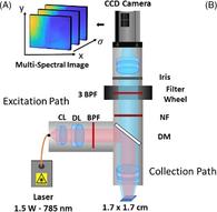
Toward noncontact macroscopic imaging of multiple cancers using multi-spectral inelastic scattering detection
Here we introduce a Raman spectroscopy approach combining multi-spectral imaging and a new fluorescence background subtraction technique to image individual Raman peaks in less than 5 seconds over a square field-of-view of 1-centimeter sides with 350 micrometers resolution. First, human data is presented supporting the feasibility of achieving cancer detection with high sensitivity and specificity – in brain, breast, lung, and ovarian/endometrium tissue – using no more than three biochemically interpretable biomarkers associated with the inelastic scattering signal from specific Raman peaks. Second, a proof-of-principle study in biological tissue is presented demonstrating the feasibility of detecting a single Raman band – here the CH2/CH3 deformation bands from proteins and lipids – using a conventional multi-spectral imaging system in combination with the new background removal method. This study paves the way for the development of a new Raman imaging technique that is rapid, label-free, and wide field.
期刊介绍:
The first international journal dedicated to publishing reviews and original articles from this exciting field, the Journal of Biophotonics covers the broad range of research on interactions between light and biological material. The journal offers a platform where the physicist communicates with the biologist and where the clinical practitioner learns about the latest tools for the diagnosis of diseases. As such, the journal is highly interdisciplinary, publishing cutting edge research in the fields of life sciences, medicine, physics, chemistry, and engineering. The coverage extends from fundamental research to specific developments, while also including the latest applications.
 求助内容:
求助内容: 应助结果提醒方式:


