{"title":"雏鸡胚胎腹腔内道的非典型发育过程","authors":"José Luis Ferran, Luis Puelles","doi":"10.1002/cne.25646","DOIUrl":null,"url":null,"abstract":"<p>Classical studies of the avian diencephalon hardly mention the habenulo-interpeduncular tract (a.k.a. retroflex tract), although both the habenula (HB) (its origin) and the interpeduncular nuclear complex (its target) are present. Retroflex tract fibers were described at early embryonic stages but seem absent in the adult in routine stains. However, this tract is a salient diencephalic landmark in all other vertebrate lineages. It typically emerges out of the caudal HB, courses dorsoventrally across thalamic alar and basal plates just in front of the thalamo-pretectal boundary, and then sharply bends 90° caudalwards at paramedian basal plate levels (this is the “retroflexion”), to approach longitudinally via paramedian pretectum and midbrain the rostralmost hindbrain, specifically the prepontine median interpeduncular complex across isthmus and rhombomere 1. We systematize this habenulo-interpeduncular course into four parts named subhabenular, retrothalamic, tegmental, and interpeduncular. We reexamined the chicken habenulo-interpeduncular fibers at stages HH30 and HH35 (6.5- and 9-day incubation) by mapping them specifically with immunoreaction for BEN protein, a well-known marker. We found that only a small fraction of the stained retroflex tract fibers approaches the basal plate by coursing along the standard dorsoventral pathway in front of the thalamo-pretectal boundary. Many other habenular fibers instead diverge into atypical dispersed courses across the thalamic cell mass (implying alteration of the first subhabenular part of the standard course) before reaching the basal plate; this dispersion explains their invisibility. A significant number of such transthalamic habenular fibers cross orthogonally the zona limitans (ZLI) (the rostral thalamic boundary) and invade the caudal alar prethalamus. Here, they immediately descend dorsoventrally, just rostrally to the ZLI, until reaching the prethalamic basal plate, where they bend (retroflex) caudalwards, entering the thalamic basal paramedian area. These atypical fibers gradually fasciculate with the other groups of habenular efferent fibers in their final longitudinal approach to the hindbrain interpeduncular complex. We conclude that the poor visibility of this tract in birds is due to its dispersion into a diversity of atypical alternative routes, though all components eventually reach the interpeduncular complex. This case merits further analysis of the diverse permissive versus nonpermissive guidance mechanisms called into action, which partially correlate distinctly with successive diencephalic, mesencephalic, and hindbrain neuromeric fields and their boundaries.</p>","PeriodicalId":15552,"journal":{"name":"Journal of Comparative Neurology","volume":"532 7","pages":""},"PeriodicalIF":2.3000,"publicationDate":"2024-07-03","publicationTypes":"Journal Article","fieldsOfStudy":null,"isOpenAccess":false,"openAccessPdf":"https://onlinelibrary.wiley.com/doi/epdf/10.1002/cne.25646","citationCount":"0","resultStr":"{\"title\":\"Atypical Course of the Habenulo-Interpeduncular Tract in Chick Embryos\",\"authors\":\"José Luis Ferran, Luis Puelles\",\"doi\":\"10.1002/cne.25646\",\"DOIUrl\":null,\"url\":null,\"abstract\":\"<p>Classical studies of the avian diencephalon hardly mention the habenulo-interpeduncular tract (a.k.a. retroflex tract), although both the habenula (HB) (its origin) and the interpeduncular nuclear complex (its target) are present. Retroflex tract fibers were described at early embryonic stages but seem absent in the adult in routine stains. However, this tract is a salient diencephalic landmark in all other vertebrate lineages. It typically emerges out of the caudal HB, courses dorsoventrally across thalamic alar and basal plates just in front of the thalamo-pretectal boundary, and then sharply bends 90° caudalwards at paramedian basal plate levels (this is the “retroflexion”), to approach longitudinally via paramedian pretectum and midbrain the rostralmost hindbrain, specifically the prepontine median interpeduncular complex across isthmus and rhombomere 1. We systematize this habenulo-interpeduncular course into four parts named subhabenular, retrothalamic, tegmental, and interpeduncular. We reexamined the chicken habenulo-interpeduncular fibers at stages HH30 and HH35 (6.5- and 9-day incubation) by mapping them specifically with immunoreaction for BEN protein, a well-known marker. We found that only a small fraction of the stained retroflex tract fibers approaches the basal plate by coursing along the standard dorsoventral pathway in front of the thalamo-pretectal boundary. Many other habenular fibers instead diverge into atypical dispersed courses across the thalamic cell mass (implying alteration of the first subhabenular part of the standard course) before reaching the basal plate; this dispersion explains their invisibility. A significant number of such transthalamic habenular fibers cross orthogonally the zona limitans (ZLI) (the rostral thalamic boundary) and invade the caudal alar prethalamus. Here, they immediately descend dorsoventrally, just rostrally to the ZLI, until reaching the prethalamic basal plate, where they bend (retroflex) caudalwards, entering the thalamic basal paramedian area. These atypical fibers gradually fasciculate with the other groups of habenular efferent fibers in their final longitudinal approach to the hindbrain interpeduncular complex. We conclude that the poor visibility of this tract in birds is due to its dispersion into a diversity of atypical alternative routes, though all components eventually reach the interpeduncular complex. This case merits further analysis of the diverse permissive versus nonpermissive guidance mechanisms called into action, which partially correlate distinctly with successive diencephalic, mesencephalic, and hindbrain neuromeric fields and their boundaries.</p>\",\"PeriodicalId\":15552,\"journal\":{\"name\":\"Journal of Comparative Neurology\",\"volume\":\"532 7\",\"pages\":\"\"},\"PeriodicalIF\":2.3000,\"publicationDate\":\"2024-07-03\",\"publicationTypes\":\"Journal Article\",\"fieldsOfStudy\":null,\"isOpenAccess\":false,\"openAccessPdf\":\"https://onlinelibrary.wiley.com/doi/epdf/10.1002/cne.25646\",\"citationCount\":\"0\",\"resultStr\":null,\"platform\":\"Semanticscholar\",\"paperid\":null,\"PeriodicalName\":\"Journal of Comparative Neurology\",\"FirstCategoryId\":\"3\",\"ListUrlMain\":\"https://onlinelibrary.wiley.com/doi/10.1002/cne.25646\",\"RegionNum\":4,\"RegionCategory\":\"医学\",\"ArticlePicture\":[],\"TitleCN\":null,\"AbstractTextCN\":null,\"PMCID\":null,\"EPubDate\":\"\",\"PubModel\":\"\",\"JCR\":\"Q3\",\"JCRName\":\"NEUROSCIENCES\",\"Score\":null,\"Total\":0}","platform":"Semanticscholar","paperid":null,"PeriodicalName":"Journal of Comparative Neurology","FirstCategoryId":"3","ListUrlMain":"https://onlinelibrary.wiley.com/doi/10.1002/cne.25646","RegionNum":4,"RegionCategory":"医学","ArticlePicture":[],"TitleCN":null,"AbstractTextCN":null,"PMCID":null,"EPubDate":"","PubModel":"","JCR":"Q3","JCRName":"NEUROSCIENCES","Score":null,"Total":0}
Atypical Course of the Habenulo-Interpeduncular Tract in Chick Embryos
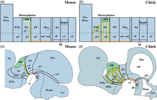
Classical studies of the avian diencephalon hardly mention the habenulo-interpeduncular tract (a.k.a. retroflex tract), although both the habenula (HB) (its origin) and the interpeduncular nuclear complex (its target) are present. Retroflex tract fibers were described at early embryonic stages but seem absent in the adult in routine stains. However, this tract is a salient diencephalic landmark in all other vertebrate lineages. It typically emerges out of the caudal HB, courses dorsoventrally across thalamic alar and basal plates just in front of the thalamo-pretectal boundary, and then sharply bends 90° caudalwards at paramedian basal plate levels (this is the “retroflexion”), to approach longitudinally via paramedian pretectum and midbrain the rostralmost hindbrain, specifically the prepontine median interpeduncular complex across isthmus and rhombomere 1. We systematize this habenulo-interpeduncular course into four parts named subhabenular, retrothalamic, tegmental, and interpeduncular. We reexamined the chicken habenulo-interpeduncular fibers at stages HH30 and HH35 (6.5- and 9-day incubation) by mapping them specifically with immunoreaction for BEN protein, a well-known marker. We found that only a small fraction of the stained retroflex tract fibers approaches the basal plate by coursing along the standard dorsoventral pathway in front of the thalamo-pretectal boundary. Many other habenular fibers instead diverge into atypical dispersed courses across the thalamic cell mass (implying alteration of the first subhabenular part of the standard course) before reaching the basal plate; this dispersion explains their invisibility. A significant number of such transthalamic habenular fibers cross orthogonally the zona limitans (ZLI) (the rostral thalamic boundary) and invade the caudal alar prethalamus. Here, they immediately descend dorsoventrally, just rostrally to the ZLI, until reaching the prethalamic basal plate, where they bend (retroflex) caudalwards, entering the thalamic basal paramedian area. These atypical fibers gradually fasciculate with the other groups of habenular efferent fibers in their final longitudinal approach to the hindbrain interpeduncular complex. We conclude that the poor visibility of this tract in birds is due to its dispersion into a diversity of atypical alternative routes, though all components eventually reach the interpeduncular complex. This case merits further analysis of the diverse permissive versus nonpermissive guidance mechanisms called into action, which partially correlate distinctly with successive diencephalic, mesencephalic, and hindbrain neuromeric fields and their boundaries.
期刊介绍:
Established in 1891, JCN is the oldest continually published basic neuroscience journal. Historically, as the name suggests, the journal focused on a comparison among species to uncover the intricacies of how the brain functions. In modern times, this research is called systems neuroscience where animal models are used to mimic core cognitive processes with the ultimate goal of understanding neural circuits and connections that give rise to behavioral patterns and different neural states.
Research published in JCN covers all species from invertebrates to humans, and the reports inform the readers about the function and organization of nervous systems in species with an emphasis on the way that species adaptations inform about the function or organization of the nervous systems, rather than on their evolution per se.
JCN publishes primary research articles and critical commentaries and review-type articles offering expert insight in to cutting edge research in the field of systems neuroscience; a complete list of contribution types is given in the Author Guidelines. For primary research contributions, only full-length investigative reports are desired; the journal does not accept short communications.

 求助内容:
求助内容: 应助结果提醒方式:


