Joshua Mayourian, Robert Geggel, William G La Cava, Sunil J Ghelani, John K Triedman
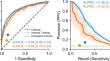
{"title":"基于深度学习的小儿心电图预测房间隔缺损","authors":"Joshua Mayourian, Robert Geggel, William G La Cava, Sunil J Ghelani, John K Triedman","doi":"10.1007/s00246-024-03540-7","DOIUrl":null,"url":null,"abstract":"<p><p>Secundum atrial septal defect (ASD2) detection is often delayed, with the potential for late diagnosis complications. Recent work demonstrated artificial intelligence-enhanced ECG analysis shows promise to detect ASD2 in adults. However, its application to pediatric populations remains underexplored. In this study, we trained a convolutional neural network (AI-pECG) on paired ECG-echocardiograms (≤ 2 days apart) to detect ASD2 from patients ≤ 18 years old without major congenital heart disease. Model performance was evaluated on the first ECG-echocardiogram pair per patient for Boston Children's Hospital internal testing and emergency department cohorts using area under the receiver operating (AUROC) and precision-recall (AUPRC) curves. The training cohort comprised of 92,377 ECG-echocardiogram pairs (46,261 patients; median age 8.2 years) with an ASD2 prevalence of 6.7%. Test groups included internal testing (12,631 patients; median age 7.4 years; 6.9% prevalence) and emergency department (2,830 patients; median age 7.5 years; 4.9% prevalence) cohorts. Model performance was higher in the internal test (AUROC 0.84, AUPRC 0.46) cohort than the emergency department cohort (AUROC 0.80, AUPRC 0.30). In both cohorts, AI-pECG outperformed ECG findings of incomplete right bundle branch block. Model explainability analyses suggest high-risk limb lead features include greater amplitude P waves (suggestive of right atrial enlargement) and V1 RSR' (suggestive of RBBB). Our findings demonstrate the promise of AI-pECG to inexpensively screen and/or detect ASD2 in pediatric patients. Future multicenter validation and prospective trials to inform clinical decision making are warranted.</p>","PeriodicalId":19814,"journal":{"name":"Pediatric Cardiology","volume":" ","pages":"1235-1240"},"PeriodicalIF":1.5000,"publicationDate":"2025-06-01","publicationTypes":"Journal Article","fieldsOfStudy":null,"isOpenAccess":false,"openAccessPdf":"https://www.ncbi.nlm.nih.gov/pmc/articles/PMC11849054/pdf/","citationCount":"0","resultStr":"{\"title\":\"Pediatric Electrocardiogram-Based Deep Learning to Predict Secundum Atrial Septal Defects.\",\"authors\":\"Joshua Mayourian, Robert Geggel, William G La Cava, Sunil J Ghelani, John K Triedman\",\"doi\":\"10.1007/s00246-024-03540-7\",\"DOIUrl\":null,\"url\":null,\"abstract\":\"<p><p>Secundum atrial septal defect (ASD2) detection is often delayed, with the potential for late diagnosis complications. Recent work demonstrated artificial intelligence-enhanced ECG analysis shows promise to detect ASD2 in adults. However, its application to pediatric populations remains underexplored. In this study, we trained a convolutional neural network (AI-pECG) on paired ECG-echocardiograms (≤ 2 days apart) to detect ASD2 from patients ≤ 18 years old without major congenital heart disease. Model performance was evaluated on the first ECG-echocardiogram pair per patient for Boston Children's Hospital internal testing and emergency department cohorts using area under the receiver operating (AUROC) and precision-recall (AUPRC) curves. The training cohort comprised of 92,377 ECG-echocardiogram pairs (46,261 patients; median age 8.2 years) with an ASD2 prevalence of 6.7%. Test groups included internal testing (12,631 patients; median age 7.4 years; 6.9% prevalence) and emergency department (2,830 patients; median age 7.5 years; 4.9% prevalence) cohorts. Model performance was higher in the internal test (AUROC 0.84, AUPRC 0.46) cohort than the emergency department cohort (AUROC 0.80, AUPRC 0.30). In both cohorts, AI-pECG outperformed ECG findings of incomplete right bundle branch block. Model explainability analyses suggest high-risk limb lead features include greater amplitude P waves (suggestive of right atrial enlargement) and V1 RSR' (suggestive of RBBB). Our findings demonstrate the promise of AI-pECG to inexpensively screen and/or detect ASD2 in pediatric patients. Future multicenter validation and prospective trials to inform clinical decision making are warranted.</p>\",\"PeriodicalId\":19814,\"journal\":{\"name\":\"Pediatric Cardiology\",\"volume\":\" \",\"pages\":\"1235-1240\"},\"PeriodicalIF\":1.5000,\"publicationDate\":\"2025-06-01\",\"publicationTypes\":\"Journal Article\",\"fieldsOfStudy\":null,\"isOpenAccess\":false,\"openAccessPdf\":\"https://www.ncbi.nlm.nih.gov/pmc/articles/PMC11849054/pdf/\",\"citationCount\":\"0\",\"resultStr\":null,\"platform\":\"Semanticscholar\",\"paperid\":null,\"PeriodicalName\":\"Pediatric Cardiology\",\"FirstCategoryId\":\"3\",\"ListUrlMain\":\"https://doi.org/10.1007/s00246-024-03540-7\",\"RegionNum\":4,\"RegionCategory\":\"医学\",\"ArticlePicture\":[],\"TitleCN\":null,\"AbstractTextCN\":null,\"PMCID\":null,\"EPubDate\":\"2024/7/2 0:00:00\",\"PubModel\":\"Epub\",\"JCR\":\"Q3\",\"JCRName\":\"CARDIAC & CARDIOVASCULAR SYSTEMS\",\"Score\":null,\"Total\":0}","platform":"Semanticscholar","paperid":null,"PeriodicalName":"Pediatric Cardiology","FirstCategoryId":"3","ListUrlMain":"https://doi.org/10.1007/s00246-024-03540-7","RegionNum":4,"RegionCategory":"医学","ArticlePicture":[],"TitleCN":null,"AbstractTextCN":null,"PMCID":null,"EPubDate":"2024/7/2 0:00:00","PubModel":"Epub","JCR":"Q3","JCRName":"CARDIAC & CARDIOVASCULAR SYSTEMS","Score":null,"Total":0}
Pediatric Electrocardiogram-Based Deep Learning to Predict Secundum Atrial Septal Defects.
Secundum atrial septal defect (ASD2) detection is often delayed, with the potential for late diagnosis complications. Recent work demonstrated artificial intelligence-enhanced ECG analysis shows promise to detect ASD2 in adults. However, its application to pediatric populations remains underexplored. In this study, we trained a convolutional neural network (AI-pECG) on paired ECG-echocardiograms (≤ 2 days apart) to detect ASD2 from patients ≤ 18 years old without major congenital heart disease. Model performance was evaluated on the first ECG-echocardiogram pair per patient for Boston Children's Hospital internal testing and emergency department cohorts using area under the receiver operating (AUROC) and precision-recall (AUPRC) curves. The training cohort comprised of 92,377 ECG-echocardiogram pairs (46,261 patients; median age 8.2 years) with an ASD2 prevalence of 6.7%. Test groups included internal testing (12,631 patients; median age 7.4 years; 6.9% prevalence) and emergency department (2,830 patients; median age 7.5 years; 4.9% prevalence) cohorts. Model performance was higher in the internal test (AUROC 0.84, AUPRC 0.46) cohort than the emergency department cohort (AUROC 0.80, AUPRC 0.30). In both cohorts, AI-pECG outperformed ECG findings of incomplete right bundle branch block. Model explainability analyses suggest high-risk limb lead features include greater amplitude P waves (suggestive of right atrial enlargement) and V1 RSR' (suggestive of RBBB). Our findings demonstrate the promise of AI-pECG to inexpensively screen and/or detect ASD2 in pediatric patients. Future multicenter validation and prospective trials to inform clinical decision making are warranted.
期刊介绍:
The editor of Pediatric Cardiology welcomes original manuscripts concerning all aspects of heart disease in infants, children, and adolescents, including embryology and anatomy, physiology and pharmacology, biochemistry, pathology, genetics, radiology, clinical aspects, investigative cardiology, electrophysiology and echocardiography, and cardiac surgery. Articles which may include original articles, review articles, letters to the editor etc., must be written in English and must be submitted solely to Pediatric Cardiology.
 求助内容:
求助内容: 应助结果提醒方式:


