{"title":"二维剪切波弹性成像和衰减成像在慢性肝病纤维化和脂肪变性评估中的诊断性能。","authors":"Tamaki Kobayashi, Takuma Nakatsuka, Masaya Sato, Yoko Soroida, Hiromi Hikita, Hiroaki Gotoh, Tomomi Iwai, Ryosuke Tateishi, Makoto Kurano, Mitsuhiro Fujishiro","doi":"10.1007/s10396-024-01473-5","DOIUrl":null,"url":null,"abstract":"<p><strong>Purpose: </strong>We investigated the diagnostic performance of two-dimensional shear wave elastography (2D-SWE) and attenuation imaging (ATI) in detecting fibrosis and steatosis in patients with chronic liver disease (CLD), comparing them with established methods.</p><p><strong>Methods: </strong>In 190 patients with CLD, 2D-SWE and vibration-controlled transient elastography (VCTE) were used for liver stiffness measurement (LSM), and ATI and controlled attenuation parameter (CAP) were used for steatosis quantification. The correlations between these new and established methods were analyzed.</p><p><strong>Results: </strong>Significant correlations were found between 2D-SWE and VCTE (r = 0.78, P < 0.001), and between ATI and CAP (r = 0.70, P < 0.001). Liver stiffness tended to be lower with 2D-SWE compared with that with VCTE, especially in cases with higher LSM, and ATI was less influenced by skin-capsular distance than CAP. Area under the receiver-operating characteristics curves (AUCs) and optimal cut-offs of 2D-SWE for diagnosing liver fibrosis stages F2, F3, and F4 were 0.73 (8.7 kPa), 0.79 (9.1 kPa), and 0.88 (11.6 kPa), respectively. The AUCs and optimal cut-offs of ATI for diagnosing hepatic steatosis grades S1, S2, and S3 were 0.91 (0.66 dB/cm/MHz), 0.80 (0.79 dB/cm/MHz), and 0.88 (0.86 dB/cm/MHz), respectively. A subgroup analysis of 86 patients with metabolic dysfunction-associated steatotic liver disease also demonstrated good performance for 2D-SWE and ATI.</p><p><strong>Conclusion: </strong>2D-SWE and ATI performed comparably with conventional VCTE and CAP in evaluating CLD, offering reliable alternatives for diagnosing liver fibrosis and steatosis.</p>","PeriodicalId":50130,"journal":{"name":"Journal of Medical Ultrasonics","volume":" ","pages":"95-103"},"PeriodicalIF":2.1000,"publicationDate":"2025-01-01","publicationTypes":"Journal Article","fieldsOfStudy":null,"isOpenAccess":false,"openAccessPdf":"https://www.ncbi.nlm.nih.gov/pmc/articles/PMC11799025/pdf/","citationCount":"0","resultStr":"{\"title\":\"Diagnostic performance of two-dimensional shear wave elastography and attenuation imaging for fibrosis and steatosis assessment in chronic liver disease.\",\"authors\":\"Tamaki Kobayashi, Takuma Nakatsuka, Masaya Sato, Yoko Soroida, Hiromi Hikita, Hiroaki Gotoh, Tomomi Iwai, Ryosuke Tateishi, Makoto Kurano, Mitsuhiro Fujishiro\",\"doi\":\"10.1007/s10396-024-01473-5\",\"DOIUrl\":null,\"url\":null,\"abstract\":\"<p><strong>Purpose: </strong>We investigated the diagnostic performance of two-dimensional shear wave elastography (2D-SWE) and attenuation imaging (ATI) in detecting fibrosis and steatosis in patients with chronic liver disease (CLD), comparing them with established methods.</p><p><strong>Methods: </strong>In 190 patients with CLD, 2D-SWE and vibration-controlled transient elastography (VCTE) were used for liver stiffness measurement (LSM), and ATI and controlled attenuation parameter (CAP) were used for steatosis quantification. The correlations between these new and established methods were analyzed.</p><p><strong>Results: </strong>Significant correlations were found between 2D-SWE and VCTE (r = 0.78, P < 0.001), and between ATI and CAP (r = 0.70, P < 0.001). Liver stiffness tended to be lower with 2D-SWE compared with that with VCTE, especially in cases with higher LSM, and ATI was less influenced by skin-capsular distance than CAP. Area under the receiver-operating characteristics curves (AUCs) and optimal cut-offs of 2D-SWE for diagnosing liver fibrosis stages F2, F3, and F4 were 0.73 (8.7 kPa), 0.79 (9.1 kPa), and 0.88 (11.6 kPa), respectively. The AUCs and optimal cut-offs of ATI for diagnosing hepatic steatosis grades S1, S2, and S3 were 0.91 (0.66 dB/cm/MHz), 0.80 (0.79 dB/cm/MHz), and 0.88 (0.86 dB/cm/MHz), respectively. A subgroup analysis of 86 patients with metabolic dysfunction-associated steatotic liver disease also demonstrated good performance for 2D-SWE and ATI.</p><p><strong>Conclusion: </strong>2D-SWE and ATI performed comparably with conventional VCTE and CAP in evaluating CLD, offering reliable alternatives for diagnosing liver fibrosis and steatosis.</p>\",\"PeriodicalId\":50130,\"journal\":{\"name\":\"Journal of Medical Ultrasonics\",\"volume\":\" \",\"pages\":\"95-103\"},\"PeriodicalIF\":2.1000,\"publicationDate\":\"2025-01-01\",\"publicationTypes\":\"Journal Article\",\"fieldsOfStudy\":null,\"isOpenAccess\":false,\"openAccessPdf\":\"https://www.ncbi.nlm.nih.gov/pmc/articles/PMC11799025/pdf/\",\"citationCount\":\"0\",\"resultStr\":null,\"platform\":\"Semanticscholar\",\"paperid\":null,\"PeriodicalName\":\"Journal of Medical Ultrasonics\",\"FirstCategoryId\":\"3\",\"ListUrlMain\":\"https://doi.org/10.1007/s10396-024-01473-5\",\"RegionNum\":4,\"RegionCategory\":\"医学\",\"ArticlePicture\":[],\"TitleCN\":null,\"AbstractTextCN\":null,\"PMCID\":null,\"EPubDate\":\"2024/6/29 0:00:00\",\"PubModel\":\"Epub\",\"JCR\":\"Q3\",\"JCRName\":\"RADIOLOGY, NUCLEAR MEDICINE & MEDICAL IMAGING\",\"Score\":null,\"Total\":0}","platform":"Semanticscholar","paperid":null,"PeriodicalName":"Journal of Medical Ultrasonics","FirstCategoryId":"3","ListUrlMain":"https://doi.org/10.1007/s10396-024-01473-5","RegionNum":4,"RegionCategory":"医学","ArticlePicture":[],"TitleCN":null,"AbstractTextCN":null,"PMCID":null,"EPubDate":"2024/6/29 0:00:00","PubModel":"Epub","JCR":"Q3","JCRName":"RADIOLOGY, NUCLEAR MEDICINE & MEDICAL IMAGING","Score":null,"Total":0}
引用次数: 0
摘要
目的:我们研究了二维剪切波弹性成像(2D-SWE)和衰减成像(ATI)在检测慢性肝病(CLD)患者肝纤维化和脂肪变性方面的诊断性能,并将其与已有方法进行了比较:在190名慢性肝病患者中,使用二维-SWE和振动控制瞬态弹性成像(VCTE)测量肝脏硬度(LSM),使用ATI和受控衰减参数(CAP)量化脂肪变性。分析了这些新方法与既有方法之间的相关性:结论:2D-SWE 和 ATI 与传统的 VCTE 和 CAP 在评估慢性肝病方面的表现相当,为诊断肝纤维化和脂肪变性提供了可靠的替代方法。
Diagnostic performance of two-dimensional shear wave elastography and attenuation imaging for fibrosis and steatosis assessment in chronic liver disease.
Purpose: We investigated the diagnostic performance of two-dimensional shear wave elastography (2D-SWE) and attenuation imaging (ATI) in detecting fibrosis and steatosis in patients with chronic liver disease (CLD), comparing them with established methods.
Methods: In 190 patients with CLD, 2D-SWE and vibration-controlled transient elastography (VCTE) were used for liver stiffness measurement (LSM), and ATI and controlled attenuation parameter (CAP) were used for steatosis quantification. The correlations between these new and established methods were analyzed.
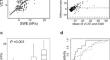
Results: Significant correlations were found between 2D-SWE and VCTE (r = 0.78, P < 0.001), and between ATI and CAP (r = 0.70, P < 0.001). Liver stiffness tended to be lower with 2D-SWE compared with that with VCTE, especially in cases with higher LSM, and ATI was less influenced by skin-capsular distance than CAP. Area under the receiver-operating characteristics curves (AUCs) and optimal cut-offs of 2D-SWE for diagnosing liver fibrosis stages F2, F3, and F4 were 0.73 (8.7 kPa), 0.79 (9.1 kPa), and 0.88 (11.6 kPa), respectively. The AUCs and optimal cut-offs of ATI for diagnosing hepatic steatosis grades S1, S2, and S3 were 0.91 (0.66 dB/cm/MHz), 0.80 (0.79 dB/cm/MHz), and 0.88 (0.86 dB/cm/MHz), respectively. A subgroup analysis of 86 patients with metabolic dysfunction-associated steatotic liver disease also demonstrated good performance for 2D-SWE and ATI.
Conclusion: 2D-SWE and ATI performed comparably with conventional VCTE and CAP in evaluating CLD, offering reliable alternatives for diagnosing liver fibrosis and steatosis.
期刊介绍:
The Journal of Medical Ultrasonics is the official journal of the Japan Society of Ultrasonics in Medicine. The main purpose of the journal is to provide forum for the publication of papers documenting recent advances and new developments in the entire field of ultrasound in medicine and biology, encompassing both the medical and the engineering aspects of the science.The journal welcomes original articles, review articles, images, and letters to the editor.The journal also provides state-of-the-art information such as announcements from the boards and the committees of the society.
 求助内容:
求助内容: 应助结果提醒方式:


