Christos Koutserimpas, Dimitrios Kotzias, Enejd Veizi, George Tsakotos, George Triantafyllou, Maria Piagkou
{"title":"探索胫骨近端形态测量中的一致比率:对膝关节置换术的启示。","authors":"Christos Koutserimpas, Dimitrios Kotzias, Enejd Veizi, George Tsakotos, George Triantafyllou, Maria Piagkou","doi":"10.1007/s00276-024-03421-x","DOIUrl":null,"url":null,"abstract":"<p><strong>Introduction: </strong>The current study, which delves into proximal tibia morphometric parameters in a Greek sample, not only analyzes whether specific linear distance ratios are consistent but also paves the way for a potential novel metric system for knee arthroplasty imaging studies using constant ratios. These findings could have significant implications for future enlarged research and clinical practice.</p><p><strong>Methods: </strong>A total of 38 dried tibiae were evaluated by two independent investigators. The following distances were measured with a digital Vernier sliding caliper: (1) the mediolateral distance of the proximal surface (A), (2) the anteroposterior distance of the proximal surface (B), (3) The longitudinal length of the bone (C), (4) the line connecting the anterior margin of the proximal surface with the highest peak of the tibia tuberosity (D), (5) the depth of the proximal margin of the medial articular facet (AF) (medial plateau) (E) and (6) the depth of the proximal margin of the lateral AF (lateral plateau) (F).</p><p><strong>Results: </strong>The A, B, C, D, E, and F mean distances were 71.3 mm, 47.4 mm, 340.2 mm, 37.1 mm, 42 mm, and 35.9 mm. Reliability analysis for each observer on all measurements revealed an interclass correlation (ICC) score of 0.975 (observer 1) and 0.971 (observer 2). The ratio A/B was 1.5, A/C was a constant 0.2, and D/C was 0.1. The ratio E/F was 1.2. The six measurements (A-F) showed excellent inter-observer reliability (all ICC values > 0.990).</p><p><strong>Conclusions: </strong>The study established constant ratios of the studied linear distances around the proximal tibia. Considering these ratios, asymmetrical tibial components in knee arthroplasty seem to replicate the native anatomy more closely. Furthermore, the distance from the anterior margin of the proximal surface to the tibial tuberosity peak, constituting one-tenth of the longitudinal length of the tibia, shows promise as a metric system for imaging studies, especially in assessing lesions around tibial components.</p>","PeriodicalId":49461,"journal":{"name":"Surgical and Radiologic Anatomy","volume":null,"pages":null},"PeriodicalIF":1.4000,"publicationDate":"2024-09-01","publicationTypes":"Journal Article","fieldsOfStudy":null,"isOpenAccess":false,"openAccessPdf":"","citationCount":"0","resultStr":"{\"title\":\"Exploring consistent ratios in morphometry of the proximal tibia: insights for knee arthroplasty.\",\"authors\":\"Christos Koutserimpas, Dimitrios Kotzias, Enejd Veizi, George Tsakotos, George Triantafyllou, Maria Piagkou\",\"doi\":\"10.1007/s00276-024-03421-x\",\"DOIUrl\":null,\"url\":null,\"abstract\":\"<p><strong>Introduction: </strong>The current study, which delves into proximal tibia morphometric parameters in a Greek sample, not only analyzes whether specific linear distance ratios are consistent but also paves the way for a potential novel metric system for knee arthroplasty imaging studies using constant ratios. These findings could have significant implications for future enlarged research and clinical practice.</p><p><strong>Methods: </strong>A total of 38 dried tibiae were evaluated by two independent investigators. The following distances were measured with a digital Vernier sliding caliper: (1) the mediolateral distance of the proximal surface (A), (2) the anteroposterior distance of the proximal surface (B), (3) The longitudinal length of the bone (C), (4) the line connecting the anterior margin of the proximal surface with the highest peak of the tibia tuberosity (D), (5) the depth of the proximal margin of the medial articular facet (AF) (medial plateau) (E) and (6) the depth of the proximal margin of the lateral AF (lateral plateau) (F).</p><p><strong>Results: </strong>The A, B, C, D, E, and F mean distances were 71.3 mm, 47.4 mm, 340.2 mm, 37.1 mm, 42 mm, and 35.9 mm. Reliability analysis for each observer on all measurements revealed an interclass correlation (ICC) score of 0.975 (observer 1) and 0.971 (observer 2). The ratio A/B was 1.5, A/C was a constant 0.2, and D/C was 0.1. The ratio E/F was 1.2. The six measurements (A-F) showed excellent inter-observer reliability (all ICC values > 0.990).</p><p><strong>Conclusions: </strong>The study established constant ratios of the studied linear distances around the proximal tibia. Considering these ratios, asymmetrical tibial components in knee arthroplasty seem to replicate the native anatomy more closely. Furthermore, the distance from the anterior margin of the proximal surface to the tibial tuberosity peak, constituting one-tenth of the longitudinal length of the tibia, shows promise as a metric system for imaging studies, especially in assessing lesions around tibial components.</p>\",\"PeriodicalId\":49461,\"journal\":{\"name\":\"Surgical and Radiologic Anatomy\",\"volume\":null,\"pages\":null},\"PeriodicalIF\":1.4000,\"publicationDate\":\"2024-09-01\",\"publicationTypes\":\"Journal Article\",\"fieldsOfStudy\":null,\"isOpenAccess\":false,\"openAccessPdf\":\"\",\"citationCount\":\"0\",\"resultStr\":null,\"platform\":\"Semanticscholar\",\"paperid\":null,\"PeriodicalName\":\"Surgical and Radiologic Anatomy\",\"FirstCategoryId\":\"3\",\"ListUrlMain\":\"https://doi.org/10.1007/s00276-024-03421-x\",\"RegionNum\":4,\"RegionCategory\":\"医学\",\"ArticlePicture\":[],\"TitleCN\":null,\"AbstractTextCN\":null,\"PMCID\":null,\"EPubDate\":\"2024/6/29 0:00:00\",\"PubModel\":\"Epub\",\"JCR\":\"Q2\",\"JCRName\":\"Medicine\",\"Score\":null,\"Total\":0}","platform":"Semanticscholar","paperid":null,"PeriodicalName":"Surgical and Radiologic Anatomy","FirstCategoryId":"3","ListUrlMain":"https://doi.org/10.1007/s00276-024-03421-x","RegionNum":4,"RegionCategory":"医学","ArticlePicture":[],"TitleCN":null,"AbstractTextCN":null,"PMCID":null,"EPubDate":"2024/6/29 0:00:00","PubModel":"Epub","JCR":"Q2","JCRName":"Medicine","Score":null,"Total":0}
Exploring consistent ratios in morphometry of the proximal tibia: insights for knee arthroplasty.
Introduction: The current study, which delves into proximal tibia morphometric parameters in a Greek sample, not only analyzes whether specific linear distance ratios are consistent but also paves the way for a potential novel metric system for knee arthroplasty imaging studies using constant ratios. These findings could have significant implications for future enlarged research and clinical practice.
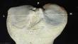
Methods: A total of 38 dried tibiae were evaluated by two independent investigators. The following distances were measured with a digital Vernier sliding caliper: (1) the mediolateral distance of the proximal surface (A), (2) the anteroposterior distance of the proximal surface (B), (3) The longitudinal length of the bone (C), (4) the line connecting the anterior margin of the proximal surface with the highest peak of the tibia tuberosity (D), (5) the depth of the proximal margin of the medial articular facet (AF) (medial plateau) (E) and (6) the depth of the proximal margin of the lateral AF (lateral plateau) (F).
Results: The A, B, C, D, E, and F mean distances were 71.3 mm, 47.4 mm, 340.2 mm, 37.1 mm, 42 mm, and 35.9 mm. Reliability analysis for each observer on all measurements revealed an interclass correlation (ICC) score of 0.975 (observer 1) and 0.971 (observer 2). The ratio A/B was 1.5, A/C was a constant 0.2, and D/C was 0.1. The ratio E/F was 1.2. The six measurements (A-F) showed excellent inter-observer reliability (all ICC values > 0.990).
Conclusions: The study established constant ratios of the studied linear distances around the proximal tibia. Considering these ratios, asymmetrical tibial components in knee arthroplasty seem to replicate the native anatomy more closely. Furthermore, the distance from the anterior margin of the proximal surface to the tibial tuberosity peak, constituting one-tenth of the longitudinal length of the tibia, shows promise as a metric system for imaging studies, especially in assessing lesions around tibial components.
期刊介绍:
Anatomy is a morphological science which cannot fail to interest the clinician. The practical application of anatomical research to clinical problems necessitates special adaptation and selectivity in choosing from numerous international works. Although there is a tendency to believe that meaningful advances in anatomy are unlikely, constant revision is necessary. Surgical and Radiologic Anatomy, the first international journal of Clinical anatomy has been created in this spirit.
Its goal is to serve clinicians, regardless of speciality-physicians, surgeons, radiologists or other specialists-as an indispensable aid with which they can improve their knowledge of anatomy. Each issue includes: Original papers, review articles, articles on the anatomical bases of medical, surgical and radiological techniques, articles of normal radiologic anatomy, brief reviews of anatomical publications of clinical interest.
Particular attention is given to high quality illustrations, which are indispensable for a better understanding of anatomical problems.
Surgical and Radiologic Anatomy is a journal written by anatomists for clinicians with a special interest in anatomy.

 求助内容:
求助内容: 应助结果提醒方式:


