R. Cicchi, E. Baria, M. Mari, G. Filippidis, D. Chorvat
{"title":"通过 GLCM 和 CT 分析从二次谐波显微镜图像中提取胶原形态特征:跨实验室研究。","authors":"R. Cicchi, E. Baria, M. Mari, G. Filippidis, D. Chorvat","doi":"10.1002/jbio.202400090","DOIUrl":null,"url":null,"abstract":"<p>Second-harmonic generation (SHG) microscopy provides a high-resolution label-free approach for noninvasively detecting collagen organization and its pathological alterations. Up to date, several imaging analysis algorithms for extracting collagen morphological features from SHG images—such as fiber size and length, order and anisotropy—have been developed. However, the dependence of extracted features on experimental setting represents a significant obstacle for translating the methodology in the clinical practice. We tackled this problem by acquiring SHG images of the same kind of collagenous sample in various laboratories using different experimental setups and imaging conditions. The acquired images were analyzed by commonly used algorithms, such as gray-level co-occurrence matrix or curvelet transform; the extracted morphological features were compared, finding that they strongly depend on some experimental parameters, whereas they are almost independent from others. We conclude with useful suggestions for comparing results obtained in different labs using different experimental setups and conditions.</p>","PeriodicalId":184,"journal":{"name":"Journal of Biophotonics","volume":"17 8","pages":""},"PeriodicalIF":2.0000,"publicationDate":"2024-06-27","publicationTypes":"Journal Article","fieldsOfStudy":null,"isOpenAccess":false,"openAccessPdf":"https://onlinelibrary.wiley.com/doi/epdf/10.1002/jbio.202400090","citationCount":"0","resultStr":"{\"title\":\"Extraction of collagen morphological features from second-harmonic generation microscopy images via GLCM and CT analyses: A cross-laboratory study\",\"authors\":\"R. Cicchi, E. Baria, M. Mari, G. Filippidis, D. Chorvat\",\"doi\":\"10.1002/jbio.202400090\",\"DOIUrl\":null,\"url\":null,\"abstract\":\"<p>Second-harmonic generation (SHG) microscopy provides a high-resolution label-free approach for noninvasively detecting collagen organization and its pathological alterations. Up to date, several imaging analysis algorithms for extracting collagen morphological features from SHG images—such as fiber size and length, order and anisotropy—have been developed. However, the dependence of extracted features on experimental setting represents a significant obstacle for translating the methodology in the clinical practice. We tackled this problem by acquiring SHG images of the same kind of collagenous sample in various laboratories using different experimental setups and imaging conditions. The acquired images were analyzed by commonly used algorithms, such as gray-level co-occurrence matrix or curvelet transform; the extracted morphological features were compared, finding that they strongly depend on some experimental parameters, whereas they are almost independent from others. We conclude with useful suggestions for comparing results obtained in different labs using different experimental setups and conditions.</p>\",\"PeriodicalId\":184,\"journal\":{\"name\":\"Journal of Biophotonics\",\"volume\":\"17 8\",\"pages\":\"\"},\"PeriodicalIF\":2.0000,\"publicationDate\":\"2024-06-27\",\"publicationTypes\":\"Journal Article\",\"fieldsOfStudy\":null,\"isOpenAccess\":false,\"openAccessPdf\":\"https://onlinelibrary.wiley.com/doi/epdf/10.1002/jbio.202400090\",\"citationCount\":\"0\",\"resultStr\":null,\"platform\":\"Semanticscholar\",\"paperid\":null,\"PeriodicalName\":\"Journal of Biophotonics\",\"FirstCategoryId\":\"101\",\"ListUrlMain\":\"https://onlinelibrary.wiley.com/doi/10.1002/jbio.202400090\",\"RegionNum\":3,\"RegionCategory\":\"物理与天体物理\",\"ArticlePicture\":[],\"TitleCN\":null,\"AbstractTextCN\":null,\"PMCID\":null,\"EPubDate\":\"\",\"PubModel\":\"\",\"JCR\":\"Q3\",\"JCRName\":\"BIOCHEMICAL RESEARCH METHODS\",\"Score\":null,\"Total\":0}","platform":"Semanticscholar","paperid":null,"PeriodicalName":"Journal of Biophotonics","FirstCategoryId":"101","ListUrlMain":"https://onlinelibrary.wiley.com/doi/10.1002/jbio.202400090","RegionNum":3,"RegionCategory":"物理与天体物理","ArticlePicture":[],"TitleCN":null,"AbstractTextCN":null,"PMCID":null,"EPubDate":"","PubModel":"","JCR":"Q3","JCRName":"BIOCHEMICAL RESEARCH METHODS","Score":null,"Total":0}
Extraction of collagen morphological features from second-harmonic generation microscopy images via GLCM and CT analyses: A cross-laboratory study
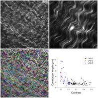
Second-harmonic generation (SHG) microscopy provides a high-resolution label-free approach for noninvasively detecting collagen organization and its pathological alterations. Up to date, several imaging analysis algorithms for extracting collagen morphological features from SHG images—such as fiber size and length, order and anisotropy—have been developed. However, the dependence of extracted features on experimental setting represents a significant obstacle for translating the methodology in the clinical practice. We tackled this problem by acquiring SHG images of the same kind of collagenous sample in various laboratories using different experimental setups and imaging conditions. The acquired images were analyzed by commonly used algorithms, such as gray-level co-occurrence matrix or curvelet transform; the extracted morphological features were compared, finding that they strongly depend on some experimental parameters, whereas they are almost independent from others. We conclude with useful suggestions for comparing results obtained in different labs using different experimental setups and conditions.
期刊介绍:
The first international journal dedicated to publishing reviews and original articles from this exciting field, the Journal of Biophotonics covers the broad range of research on interactions between light and biological material. The journal offers a platform where the physicist communicates with the biologist and where the clinical practitioner learns about the latest tools for the diagnosis of diseases. As such, the journal is highly interdisciplinary, publishing cutting edge research in the fields of life sciences, medicine, physics, chemistry, and engineering. The coverage extends from fundamental research to specific developments, while also including the latest applications.
 求助内容:
求助内容: 应助结果提醒方式:


