C Zhang, L Zhang, J X Ren, J Y Li, L P Tan, L Gao, C J Zhao
{"title":"[白细胞介素-22 通过调节微生物群和 E-cadherin 表达保护牙龈上皮屏障的研究]。","authors":"C Zhang, L Zhang, J X Ren, J Y Li, L P Tan, L Gao, C J Zhao","doi":"10.3760/cma.j.cn112144-20231115-00252","DOIUrl":null,"url":null,"abstract":"<p><p><b>Objective:</b> To investigate the regulatory effect and mechanism of interleukin-22 (IL-22) on the gingival epithelial barrier in the context of periodontal inflammation. <b>Methods:</b> IL-22 knockout (IL-22 KO) mice were constructed, and periodontitis mice models were established through oral gavage with polymicrobial inoculation. DNAs were extracted from the oral plaques of IL-22 KO periodontitis mice group (<i>n=</i>7) and their wild-type littermates periodontitis group (<i>n=</i>7) to establish a periodontitis-related oral microbiota database\"PD-RiskMicroDB\", determining the relationship between changes in oral microbiota and microbial function in two groups using 16S rRNA sequencing results. Gingival epithelial cells (GEC) were cultured by modified trypsinization method, and were stimulated with 100 μg/L IL-22, <i>Porphyromonas gingivalis</i> (Pg) (multiplicity of infection:100), separately or together for 3 and 12 hours. The experimental groups were as follows: control group (no stimulation), IL-22 group, Pg group and Pg+IL-22 group. The expression of barrier protein E-cadherin in each group at 3 h was detected by immunofluorescence, real-time fluorescence quantitative PCR (RT-qPCR) and Western blotting. Fluorescein isothiocyanate-dextran-mediated epithelial cell permeability experiment was conducted to clarify the changes in permeability of GEC in each group at 3 and 12 h. The mRNA expressions of E-cadherin in the gingival epithelium of wild-type littermates periodontitis group and IL-22 KO periodontitis group were detected by RT-qPCR. Fifteen C57BL/6 wild-type mice were randomly divided into control group (<i>n=</i>5), periodontitis group (<i>n=</i>5) and periodontitis+IL-22 treatment group (<i>n=</i>5). RT-qPCR and immunohistochemistry (IHC) staining were used to detect the expression level of E-cadherin in the gingival epithelium of each group. <b>Results:</b> 16S rRNA sequencing results showed that the composition of oral microbiota changed in IL-22 KO periodontitis group, of which the abundance of bacterial genera related to periodontal tissue invasion was significantly increased (linear discriminant analysis score: 2.22, <i>P=</i>0.009), compared with wild-type littermates periodontitis group. <i>In vitro</i> cell experiments showed that after Pg infection for 3 hours, the cell connections of GEC in Pg group were interrupted, and the fluorescence intensity of E-cadherin was reduced in Pg group compared with the control group. Meanwhile, the mRNA and protein expression levels of E-cadherin (mRNA: 0.69±0.12; protein: 0.60±0.12) were downregulated compared with the control group [mRNA: 1.00±0.00 (<i>P=</i>0.043); protein: 1.04±0.08 (<i>P=</i>0.003)], respectively. The fluorescence intensity of E-cadherin in the Pg+IL-22 group was enhanced compared with Pg group, and expression levels of E-cadherin mRNA (1.16±0.10) and protein (0.98±0.07) in Pg+IL-22 group showed a significant increase compared with Pg group [mRNA: 0.69±0.12 (<i>P=</i>0.005); protein: 0.60±0.12 (<i>P=</i>0.007)]. The result of epithelial permeability test showed that there was no statistical difference in epithelial permeability among control group, Pg group, IL-22 group and Pg+IL-22 group with treatment for 3 hours (<i>F=</i>0.20, <i>P=</i>0.893). While when the treatment time turned to be 12 hours, the epithelial barrier permeability showed a significant increase in Pg group (1.39±0.15) compared with control group (1.00±0.00, <i>P=</i>0.027), and a decrease in Pg+IL-22 group (1.02±0.18) compared with Pg group (1.39±0.15, <i>P=</i>0.034). <i>In vivo</i>, the mRNA expression of E-cadherin in the gingival epithelium of IL-22 KO periodontitis group decreased significantly (0.32±0.21) compared with wild-type littermates periodontitis group (1.01±0.01) (<i>t=</i>5.70, <i>P=</i>0.005). Moreover, RT-qPCR and IHC staining results showed that the mRNA expression level of E-cadherin (0.40±0.07) and absorbance value of E-cadherin positive expression (0.02±0.00) in gingival epithelial tissue of periodontitis group were both significantly down-regulated compared with control group [mRNA: 1.00±0.00 (<i>P=</i>0.005); absorbance value of E-cadherin positive expression: 0.04±0.01 (<i>P=</i>0.006)]. Meanwhile, the mRNA expression level of E-cadherin (1.06±0.24) and the absorbance value of E-cadherin positive expression (0.03±0.01) were both observed increase in periodontitis+IL-22 treatment group compared with periodontitis group (<i>P=</i>0.003, <i>P=</i>0.039). <b>Conclusions:</b> IL-22 may exert a protective effect on the gingival epithelial barrier in an inflammatory environment by regulating the invasiveness of oral microbiota and the expression of host barrier protein.</p>","PeriodicalId":23965,"journal":{"name":"中华口腔医学杂志","volume":"59 7","pages":"653-662"},"PeriodicalIF":0.0000,"publicationDate":"2024-07-09","publicationTypes":"Journal Article","fieldsOfStudy":null,"isOpenAccess":false,"openAccessPdf":"","citationCount":"0","resultStr":"{\"title\":\"[Study on the protection of gingival epithelial barrier by interleukin-22 through regulating microbiota and E-cadherin expression].\",\"authors\":\"C Zhang, L Zhang, J X Ren, J Y Li, L P Tan, L Gao, C J Zhao\",\"doi\":\"10.3760/cma.j.cn112144-20231115-00252\",\"DOIUrl\":null,\"url\":null,\"abstract\":\"<p><p><b>Objective:</b> To investigate the regulatory effect and mechanism of interleukin-22 (IL-22) on the gingival epithelial barrier in the context of periodontal inflammation. <b>Methods:</b> IL-22 knockout (IL-22 KO) mice were constructed, and periodontitis mice models were established through oral gavage with polymicrobial inoculation. DNAs were extracted from the oral plaques of IL-22 KO periodontitis mice group (<i>n=</i>7) and their wild-type littermates periodontitis group (<i>n=</i>7) to establish a periodontitis-related oral microbiota database\\\"PD-RiskMicroDB\\\", determining the relationship between changes in oral microbiota and microbial function in two groups using 16S rRNA sequencing results. Gingival epithelial cells (GEC) were cultured by modified trypsinization method, and were stimulated with 100 μg/L IL-22, <i>Porphyromonas gingivalis</i> (Pg) (multiplicity of infection:100), separately or together for 3 and 12 hours. The experimental groups were as follows: control group (no stimulation), IL-22 group, Pg group and Pg+IL-22 group. The expression of barrier protein E-cadherin in each group at 3 h was detected by immunofluorescence, real-time fluorescence quantitative PCR (RT-qPCR) and Western blotting. Fluorescein isothiocyanate-dextran-mediated epithelial cell permeability experiment was conducted to clarify the changes in permeability of GEC in each group at 3 and 12 h. The mRNA expressions of E-cadherin in the gingival epithelium of wild-type littermates periodontitis group and IL-22 KO periodontitis group were detected by RT-qPCR. Fifteen C57BL/6 wild-type mice were randomly divided into control group (<i>n=</i>5), periodontitis group (<i>n=</i>5) and periodontitis+IL-22 treatment group (<i>n=</i>5). RT-qPCR and immunohistochemistry (IHC) staining were used to detect the expression level of E-cadherin in the gingival epithelium of each group. <b>Results:</b> 16S rRNA sequencing results showed that the composition of oral microbiota changed in IL-22 KO periodontitis group, of which the abundance of bacterial genera related to periodontal tissue invasion was significantly increased (linear discriminant analysis score: 2.22, <i>P=</i>0.009), compared with wild-type littermates periodontitis group. <i>In vitro</i> cell experiments showed that after Pg infection for 3 hours, the cell connections of GEC in Pg group were interrupted, and the fluorescence intensity of E-cadherin was reduced in Pg group compared with the control group. Meanwhile, the mRNA and protein expression levels of E-cadherin (mRNA: 0.69±0.12; protein: 0.60±0.12) were downregulated compared with the control group [mRNA: 1.00±0.00 (<i>P=</i>0.043); protein: 1.04±0.08 (<i>P=</i>0.003)], respectively. The fluorescence intensity of E-cadherin in the Pg+IL-22 group was enhanced compared with Pg group, and expression levels of E-cadherin mRNA (1.16±0.10) and protein (0.98±0.07) in Pg+IL-22 group showed a significant increase compared with Pg group [mRNA: 0.69±0.12 (<i>P=</i>0.005); protein: 0.60±0.12 (<i>P=</i>0.007)]. The result of epithelial permeability test showed that there was no statistical difference in epithelial permeability among control group, Pg group, IL-22 group and Pg+IL-22 group with treatment for 3 hours (<i>F=</i>0.20, <i>P=</i>0.893). While when the treatment time turned to be 12 hours, the epithelial barrier permeability showed a significant increase in Pg group (1.39±0.15) compared with control group (1.00±0.00, <i>P=</i>0.027), and a decrease in Pg+IL-22 group (1.02±0.18) compared with Pg group (1.39±0.15, <i>P=</i>0.034). <i>In vivo</i>, the mRNA expression of E-cadherin in the gingival epithelium of IL-22 KO periodontitis group decreased significantly (0.32±0.21) compared with wild-type littermates periodontitis group (1.01±0.01) (<i>t=</i>5.70, <i>P=</i>0.005). Moreover, RT-qPCR and IHC staining results showed that the mRNA expression level of E-cadherin (0.40±0.07) and absorbance value of E-cadherin positive expression (0.02±0.00) in gingival epithelial tissue of periodontitis group were both significantly down-regulated compared with control group [mRNA: 1.00±0.00 (<i>P=</i>0.005); absorbance value of E-cadherin positive expression: 0.04±0.01 (<i>P=</i>0.006)]. Meanwhile, the mRNA expression level of E-cadherin (1.06±0.24) and the absorbance value of E-cadherin positive expression (0.03±0.01) were both observed increase in periodontitis+IL-22 treatment group compared with periodontitis group (<i>P=</i>0.003, <i>P=</i>0.039). <b>Conclusions:</b> IL-22 may exert a protective effect on the gingival epithelial barrier in an inflammatory environment by regulating the invasiveness of oral microbiota and the expression of host barrier protein.</p>\",\"PeriodicalId\":23965,\"journal\":{\"name\":\"中华口腔医学杂志\",\"volume\":\"59 7\",\"pages\":\"653-662\"},\"PeriodicalIF\":0.0000,\"publicationDate\":\"2024-07-09\",\"publicationTypes\":\"Journal Article\",\"fieldsOfStudy\":null,\"isOpenAccess\":false,\"openAccessPdf\":\"\",\"citationCount\":\"0\",\"resultStr\":null,\"platform\":\"Semanticscholar\",\"paperid\":null,\"PeriodicalName\":\"中华口腔医学杂志\",\"FirstCategoryId\":\"3\",\"ListUrlMain\":\"https://doi.org/10.3760/cma.j.cn112144-20231115-00252\",\"RegionNum\":0,\"RegionCategory\":null,\"ArticlePicture\":[],\"TitleCN\":null,\"AbstractTextCN\":null,\"PMCID\":null,\"EPubDate\":\"\",\"PubModel\":\"\",\"JCR\":\"Q4\",\"JCRName\":\"Medicine\",\"Score\":null,\"Total\":0}","platform":"Semanticscholar","paperid":null,"PeriodicalName":"中华口腔医学杂志","FirstCategoryId":"3","ListUrlMain":"https://doi.org/10.3760/cma.j.cn112144-20231115-00252","RegionNum":0,"RegionCategory":null,"ArticlePicture":[],"TitleCN":null,"AbstractTextCN":null,"PMCID":null,"EPubDate":"","PubModel":"","JCR":"Q4","JCRName":"Medicine","Score":null,"Total":0}
[Study on the protection of gingival epithelial barrier by interleukin-22 through regulating microbiota and E-cadherin expression].
Objective: To investigate the regulatory effect and mechanism of interleukin-22 (IL-22) on the gingival epithelial barrier in the context of periodontal inflammation. Methods: IL-22 knockout (IL-22 KO) mice were constructed, and periodontitis mice models were established through oral gavage with polymicrobial inoculation. DNAs were extracted from the oral plaques of IL-22 KO periodontitis mice group (n=7) and their wild-type littermates periodontitis group (n=7) to establish a periodontitis-related oral microbiota database"PD-RiskMicroDB", determining the relationship between changes in oral microbiota and microbial function in two groups using 16S rRNA sequencing results. Gingival epithelial cells (GEC) were cultured by modified trypsinization method, and were stimulated with 100 μg/L IL-22, Porphyromonas gingivalis (Pg) (multiplicity of infection:100), separately or together for 3 and 12 hours. The experimental groups were as follows: control group (no stimulation), IL-22 group, Pg group and Pg+IL-22 group. The expression of barrier protein E-cadherin in each group at 3 h was detected by immunofluorescence, real-time fluorescence quantitative PCR (RT-qPCR) and Western blotting. Fluorescein isothiocyanate-dextran-mediated epithelial cell permeability experiment was conducted to clarify the changes in permeability of GEC in each group at 3 and 12 h. The mRNA expressions of E-cadherin in the gingival epithelium of wild-type littermates periodontitis group and IL-22 KO periodontitis group were detected by RT-qPCR. Fifteen C57BL/6 wild-type mice were randomly divided into control group (n=5), periodontitis group (n=5) and periodontitis+IL-22 treatment group (n=5). RT-qPCR and immunohistochemistry (IHC) staining were used to detect the expression level of E-cadherin in the gingival epithelium of each group. Results: 16S rRNA sequencing results showed that the composition of oral microbiota changed in IL-22 KO periodontitis group, of which the abundance of bacterial genera related to periodontal tissue invasion was significantly increased (linear discriminant analysis score: 2.22, P=0.009), compared with wild-type littermates periodontitis group. In vitro cell experiments showed that after Pg infection for 3 hours, the cell connections of GEC in Pg group were interrupted, and the fluorescence intensity of E-cadherin was reduced in Pg group compared with the control group. Meanwhile, the mRNA and protein expression levels of E-cadherin (mRNA: 0.69±0.12; protein: 0.60±0.12) were downregulated compared with the control group [mRNA: 1.00±0.00 (P=0.043); protein: 1.04±0.08 (P=0.003)], respectively. The fluorescence intensity of E-cadherin in the Pg+IL-22 group was enhanced compared with Pg group, and expression levels of E-cadherin mRNA (1.16±0.10) and protein (0.98±0.07) in Pg+IL-22 group showed a significant increase compared with Pg group [mRNA: 0.69±0.12 (P=0.005); protein: 0.60±0.12 (P=0.007)]. The result of epithelial permeability test showed that there was no statistical difference in epithelial permeability among control group, Pg group, IL-22 group and Pg+IL-22 group with treatment for 3 hours (F=0.20, P=0.893). While when the treatment time turned to be 12 hours, the epithelial barrier permeability showed a significant increase in Pg group (1.39±0.15) compared with control group (1.00±0.00, P=0.027), and a decrease in Pg+IL-22 group (1.02±0.18) compared with Pg group (1.39±0.15, P=0.034). In vivo, the mRNA expression of E-cadherin in the gingival epithelium of IL-22 KO periodontitis group decreased significantly (0.32±0.21) compared with wild-type littermates periodontitis group (1.01±0.01) (t=5.70, P=0.005). Moreover, RT-qPCR and IHC staining results showed that the mRNA expression level of E-cadherin (0.40±0.07) and absorbance value of E-cadherin positive expression (0.02±0.00) in gingival epithelial tissue of periodontitis group were both significantly down-regulated compared with control group [mRNA: 1.00±0.00 (P=0.005); absorbance value of E-cadherin positive expression: 0.04±0.01 (P=0.006)]. Meanwhile, the mRNA expression level of E-cadherin (1.06±0.24) and the absorbance value of E-cadherin positive expression (0.03±0.01) were both observed increase in periodontitis+IL-22 treatment group compared with periodontitis group (P=0.003, P=0.039). Conclusions: IL-22 may exert a protective effect on the gingival epithelial barrier in an inflammatory environment by regulating the invasiveness of oral microbiota and the expression of host barrier protein.
期刊介绍:
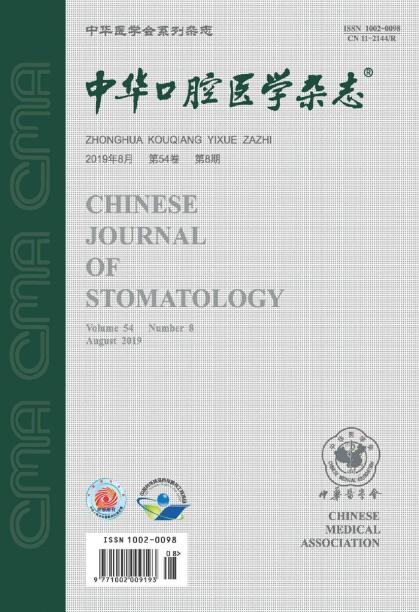
Founded in August 1953, Chinese Journal of Stomatology is a monthly academic journal of stomatology published publicly at home and abroad, sponsored by the Chinese Medical Association and co-sponsored by the Chinese Stomatology Association. It mainly reports the leading scientific research results and clinical diagnosis and treatment experience in the field of oral medicine, as well as the basic theoretical research that has a guiding role in oral clinical practice and is closely combined with oral clinical practice.
Chinese Journal of Over the years, Stomatology has been published in Medline, Scopus database, Toxicology Abstracts Database, Chemical Abstracts Database, American Cancer database, Russian Abstracts database, China Core Journal of Science and Technology, Peking University Core Journal, CSCD and other more than 20 important journals at home and abroad Physical medicine database and retrieval system included.