Dimitrios Chatzakis, Roula Al Belbeisi, Soultana Karagianni, Eustratios Karagiannakidis, George Koumoundouros
{"title":"东地中海鱼类骨骼异常的发生率和类型。","authors":"Dimitrios Chatzakis, Roula Al Belbeisi, Soultana Karagianni, Eustratios Karagiannakidis, George Koumoundouros","doi":"10.1111/jfd.13992","DOIUrl":null,"url":null,"abstract":"<p>The development of morpho-anatomical abnormalities in fish was first figured in the 16th and 17th centuries (1555–1642, Gudger, <span>1936</span>). In his extensive literature review, Dawson (<span>1964</span>) reported 1020 publications on the presence of morpho-anatomical abnormalities (including skeletal, non-skeletal, and pigmentation defects) in wild fish. A few years later, the same author updated this literature list with 213 new publications, 137 of which were published between 1965 and 1970 (Dawson, <span>1971</span>). Since then, reports on the presence of skeletal abnormalities in wild fish populations are getting more frequent. This might be attributed to the effects of anthropogenic pressures (e.g., pollution, eutrophication) on the stocks (Boglione, <span>2020</span>; Diggles, <span>2013</span>; García-Gasca et al., <span>2016</span>; Leone et al., <span>2021</span>; Messaoudi, Deli, et al., <span>2009</span>), to the exponential increase of scientific awareness about this issue, as well as to the increasing amount of research on bone deformities in aquaculture and the significant research being undertaken to understand the causes (e.g., Boglione et al., <span>2013</span>; Ytteborg et al., <span>2012</span>). Generally, the literature on deformed fish is prevailed by records of one to a few individuals occasionally observed in the field (e.g., Grimaldi & Bertoncini, <span>2021</span>; Jawad & Ibrahim, <span>2018a</span>; Yamamoto et al., <span>2013</span>). Existing studies document that these rates may elevate to high levels, varying among the species (e.g., 11% in <i>Labrus bergylta</i> vs. 57% in <i>Cyclopterus lumpus</i>, Fjelldal et al., <span>2021</span>; 9% in <i>Liza aurata</i> vs. 21% in <i>Chelon labrosus</i>, Boglione et al., <span>2006</span>), and the study area (e.g., 29% in polluted vs 3% in unpolluted sites, <i>Aphanius fasciatus</i>, Kessabi, Annabi, et al., <span>2013</span>; Kessabi, Said, & Messaoudi, <span>2013</span>; 12%–16% in polluted vs. 2%–9% in unpolluted estuaries, García-Gasca et al., <span>2016</span>). Studies on reared fish demonstrate that skeletal abnormalities have significant adverse effects on fish form and function (i.e., growth and survival rates, susceptibility to diseases) (reviewed by Boglione et al., <span>2013</span>). Since these effects are expected to be magnified in the wild, skeletal abnormalities may constitute a significant factor of natural mortality and present a decreasing prevalence with fish age (Kessabi et al., <span>2009</span>; Pollock, <span>2015</span>).</p><p>The status of abnormality prevalence in the wild fish of the Mediterranean Sea remains unknown. As a result, there are no reference data for future monitoring programs and the effect of fish abnormalities on stock natural mortality cannot be estimated. Existing studies on skeletal abnormalities in Mediterranean fishes have primarily focused on the use of fish abnormalities as bioindicators to assess the environmental quality of specific Italian lagoons (Boglione et al., <span>2006</span>; Leone et al., <span>2021</span>) and polluted areas in the Gulf of Gabes in Tunisia (Kessabi et al., <span>2009</span>; Kessabi, Annabi, et al., <span>2013</span>; Kessabi, Said, & Messaoudi, <span>2013</span>; Messaoudi, Deli, et al., <span>2009</span>; Messaoudi, Kessabi, et al., <span>2009</span>). In each of these cases, the results consistently endorse the feasibility of monitoring abnormalities in fish as a viable method for promptly identifying anthropogenic influences in the Mediterranean Sea.</p><p>In the present study, we report the presence of different types of skeletal abnormalities in specimens of 10 fish species from the Aegean Sea. Estimations of abnormality prevalence are provided for seven species, whereas for three species we provide only single abnormality records. Fish samples were collected in Chania area (Crete, South Aegean Sea, Greece, Figure 1), both from recreational and commercial small-scale fisheries. Fish were transferred to the laboratory and digital images of the left side of each individual were captured. All specimens were then stored at −20°C (Laboratory of Marine Biology, Biology Department, University of Crete). Fish radiography was performed by using a Mex + 60 x-ray generator (medical ECONET GmbH, Oberhausen, Germany, 40–44 kV, 1.0–1.2 mA s, 75 cm focal distance) and a Dura-line XD 14 wireless DR detector (AGFA, Mortsel, Belgium).</p><p>A total of nine types of skeletal abnormalities were detected on jaws, vertebral column, dorsal and caudal fins, at rates of 3.3%–28.6% (Table 1). Vertebral abnormalities consisted of lordosis (V-shape bending of the vertebral column), kyphosis (Λ-shape bending of the vertebral column) and kypholordosis (concurrent lordosis and kyphosis) (Figures 2 and 3). In the studied <i>Boops boops</i> (Linnaeus, 1758) specimens, vertebral abnormalities consisted exclusively of kypholordosis (4.2%–10.3%), without any deviation from the normal meristic counts of the vertebrae and their processes (24 vertebrae) (Figure 2a,b). A single specimen of <i>Mullus surmuletus</i> (Linnaeus, 1758) presented concurrently three lordotic areas along its vertebral column (Figure 2c). Axis abnormalities in <i>Sparisoma cretense</i> (Linnaeus, 1758) (3.7%, Table 1) consisted of kyphosis of the haemal vertebral area, which was accompanied by an abnormally straightened anteriormost part of the axis, an upward shift of the skull (stargazer) and depressions of the dorsal profile of the body (Figure 2d,d′,e). A similar upward shift of the skull and depressions of the dorsal profile were observed in <i>Pterois miles</i> (Bennett, 1828) with lordosis (4.0%, Table 1) of the causal peduncle (Figure 3a,b). In this specimen, lordosis was associated with a fusion of the pre-ural centra 2 and 3 (PU2, PU3) into one bearing two haemal processes (Figure 3a′,b′). Finally, a <i>Serranus scriba</i> (Bennett, 1828) specimen (3.3%, Table 1) was recorded to present lordosis of the prehaemal area, linked with an upward shift of the skull (stargazer) (Figure 3c,d). Of the vertebral abnormalities recorded in the present study, kypholordosis is probably the most frequently reported in the wild fish (e.g., Bengtsson et al., <span>1985</span>; Bensaada et al., <span>2023</span>; Jawad et al., <span>2017</span>; Jawad & Ibrahim, <span>2018a</span>; Mariasingarayan et al., <span>2022</span>; Van Den Avyle et al., <span>1989</span>). On the other hand, to our knowledge, this is the first record of severe lordosis of the caudal peduncle, and of the abnormally straightened anteriormost part of the axis in natural fish populations.</p><p>In the present study, recorded skull abnormalities consisted of the compression of the ethmoid area and upper jaws (pugheadedness), shortened lower jaw, missing premaxillaries and lateral misalignment of the upper and lower jaws (crossbite). The highest frequency of pugheadedness was observed in one <i>Diplodus sargus</i> (Linnaeus, 1758) sample (28.6%, Table 1, Figure 4a–c). Pugheadedness and shortened lower jaw were present in both the examined samples of <i>Pagellus erythrinus</i> (Linnaeus, 1758) (Figure 4d–g), with equal frequencies (1.8%–2.8% for each abnormality type, Table 1). In one <i>P. erythrinus</i> specimen, pugheadedness was accompanied by crossbite (Figure 4e,e′). Missing premaxillaries were observed in one kypholordotic specimen of <i>B. boops</i> (Figure 4h). Of the skull abnormalities recorded in the present study, pugheadedness is the most frequently reported in wild fish (Bueno et al., <span>2015</span>; Jawad et al., <span>2017</span>; Näslund & Jawad, <span>2022</span>; Porta & Snow, <span>2019</span>). To our knowledge, crossbite and shortened lower jaw are frequently reported to develop in reared (Boglione et al., <span>2013</span>; Fragkoulis et al., <span>2018</span>), but rarely in wild fish.</p><p>In the present study, recorded fin abnormalities mainly consisted of the partial-to-complete lack of the dorsal fin, and/or depressions of the dorsal profile of the body (saddleback syndrome) (four species, Table 1). In one specimen of <i>Siganus rivulatus</i> Forsskål & Niebuhr (1775), the depression of the dorsal profile of the body was not associated with missing rays, but with a kyphosis of the adjacent vertebrae and abnormally shaped neural processes (Figure 5a,a′). In the second abnormal specimen of <i>S. rivulatus</i> detected, as well as in one <i>S. cretense</i> specimen, a dorsal body depression was associated with abnormal adjacent pterygiophores and missing rays (Figure 5b,b′,c,c′). A single specimen of <i>D. sargus</i> presented an anterior dorsal depression of the body, which was anatomically associated with abnormal shape and position of the predorsal elements (Figure 5d,d″). Finally, a single specimen of <i>Symphodus tinca</i> (Linnaeus, 1758) and <i>Solea solea</i> (Linnaeus, 1758) presented an entire missing part of its dorsal body area (Figure 5e,e′) and an entirely missing caudal fin (Figure 5f,f′) respectively. The term “saddleback syndrome” (SBS) was initially introduced by Tave et al. (<span>1983</span>) to characterize the partial-to-complete absence of the dorsal fin in tilapia <i>Sarotherodon aureus</i>. Since its inception, SBS has been observed to manifest in various fish species under captive conditions (Boglione et al., <span>2003</span>; Cobcroft & Battaglene, <span>2013</span>; Fragkoulis et al., <span>2017</span>; Koumoundouros et al., <span>2001</span>) and in wild fish populations globally (e.g., in 10 species by Browder et al., <span>1993</span>; in <i>S. cretense</i> by Koumoundouros, <span>2008</span>; in <i>Acanthopagrus australis</i> by Diggles, <span>2013</span>; Pollock, <span>2015</span>; in 10 species by Jawad & Ibrahim, <span>2018b</span>; in <i>Parastromateus niger</i> by Silambarasan et al., <span>2021</span>; in <i>Lates calcarifer</i> by Abed et al., <span>2023</span>). Notably, Diggles (<span>2013</span>) documented an approximately sixfold increase in the prevalence of SBS among wild <i>A. australis</i> over a span of 20 years. The missing part of dorsal profile in <i>S. tinca</i> (present study) is similar to the abnormality reported in <i>P. miles</i>, which was attributed to physical injuries related to failed spearfishing attempts (Jimenez et al., <span>2022</span>).</p><p>Skeletal abnormalities in fish may be induced by unfavourable environmental and nutritional conditions (reviewed by Boglione et al., <span>2013</span>), genetic factors (e.g., Fragkoulis et al., <span>2020</span>; Takeuchi, <span>1966</span>; Tave et al., <span>1983</span>), diseases (Ngo et al., <span>2024</span>; Pasnik et al., <span>2007</span>; Piamsomboon et al., <span>2022</span>), pollutants (e.g., Johnson et al., <span>2020</span>; Kessabi, Annabi, et al., <span>2013</span>; Kessabi, Said, & Messaoudi, <span>2013</span>; Messaoudi, Deli, et al., <span>2009</span>), or toxins from harmful algal blooms (Sergi et al., <span>2022</span>). In the present study, differences in abnormality rates and types among the studied species might be related to differences in life mode, feeding habits, reproduction pattern and seasonality. For example, <i>B. boops</i> juveniles migrate to the anthropogenically modified littoral zone (e.g., small harbours) (Georgiadis et al., <span>2014</span>), where they may be subjected to various environmental stressors. A substantial number of abnormal specimens was collected by the present study in two small marinas. The high abnormality rates observed in the case of kypholordosis in <i>B. boops</i> (10.3%) and pugheadedness in <i>D. sargus</i> (28.6%) (Table 1), might be the result of localized causative factors, but also of the aggregation of deformed individuals in a protected environment where predation pressure is lower than in the open sea.</p><p>Publications reporting the presence of skeletal abnormalities in wild fish have been increasing in recent years. Since several of these abnormalities have significant adverse effects on fish performance (e.g., swimming, feeding), they could be considered as an additional source of mortality for natural stocks. However, the current available information does not allow for an estimation of the magnitude of the problem. This is mainly due to the lack of relevant monitoring surveys conducted at both spatial and temporal levels, which should also involve sampling at different developmental periods.</p><p>\n <b>Dimitrios Chatzakis:</b> Investigation; formal analysis. <b>Roula Al Belbeisi:</b> Investigation; formal analysis. <b>Soultana Karagianni:</b> Investigation; formal analysis. <b>Eustratios Karagiannakidis:</b> Visualization; methodology. <b>George Koumoundouros:</b> Formal analysis; writing – review and editing; writing – original draft; conceptualization; supervision.</p><p>This study was funded by the Property Development and Management Company of the University of Crete (Project no: 98).</p><p>The authors declare no conflict of interest.</p>","PeriodicalId":15849,"journal":{"name":"Journal of fish diseases","volume":"47 10","pages":""},"PeriodicalIF":2.2000,"publicationDate":"2024-06-29","publicationTypes":"Journal Article","fieldsOfStudy":null,"isOpenAccess":false,"openAccessPdf":"https://onlinelibrary.wiley.com/doi/epdf/10.1111/jfd.13992","citationCount":"0","resultStr":"{\"title\":\"Prevalence and typology of skeletal abnormalities in fishes of the Eastern Mediterranean\",\"authors\":\"Dimitrios Chatzakis, Roula Al Belbeisi, Soultana Karagianni, Eustratios Karagiannakidis, George Koumoundouros\",\"doi\":\"10.1111/jfd.13992\",\"DOIUrl\":null,\"url\":null,\"abstract\":\"<p>The development of morpho-anatomical abnormalities in fish was first figured in the 16th and 17th centuries (1555–1642, Gudger, <span>1936</span>). In his extensive literature review, Dawson (<span>1964</span>) reported 1020 publications on the presence of morpho-anatomical abnormalities (including skeletal, non-skeletal, and pigmentation defects) in wild fish. A few years later, the same author updated this literature list with 213 new publications, 137 of which were published between 1965 and 1970 (Dawson, <span>1971</span>). Since then, reports on the presence of skeletal abnormalities in wild fish populations are getting more frequent. This might be attributed to the effects of anthropogenic pressures (e.g., pollution, eutrophication) on the stocks (Boglione, <span>2020</span>; Diggles, <span>2013</span>; García-Gasca et al., <span>2016</span>; Leone et al., <span>2021</span>; Messaoudi, Deli, et al., <span>2009</span>), to the exponential increase of scientific awareness about this issue, as well as to the increasing amount of research on bone deformities in aquaculture and the significant research being undertaken to understand the causes (e.g., Boglione et al., <span>2013</span>; Ytteborg et al., <span>2012</span>). Generally, the literature on deformed fish is prevailed by records of one to a few individuals occasionally observed in the field (e.g., Grimaldi & Bertoncini, <span>2021</span>; Jawad & Ibrahim, <span>2018a</span>; Yamamoto et al., <span>2013</span>). Existing studies document that these rates may elevate to high levels, varying among the species (e.g., 11% in <i>Labrus bergylta</i> vs. 57% in <i>Cyclopterus lumpus</i>, Fjelldal et al., <span>2021</span>; 9% in <i>Liza aurata</i> vs. 21% in <i>Chelon labrosus</i>, Boglione et al., <span>2006</span>), and the study area (e.g., 29% in polluted vs 3% in unpolluted sites, <i>Aphanius fasciatus</i>, Kessabi, Annabi, et al., <span>2013</span>; Kessabi, Said, & Messaoudi, <span>2013</span>; 12%–16% in polluted vs. 2%–9% in unpolluted estuaries, García-Gasca et al., <span>2016</span>). Studies on reared fish demonstrate that skeletal abnormalities have significant adverse effects on fish form and function (i.e., growth and survival rates, susceptibility to diseases) (reviewed by Boglione et al., <span>2013</span>). Since these effects are expected to be magnified in the wild, skeletal abnormalities may constitute a significant factor of natural mortality and present a decreasing prevalence with fish age (Kessabi et al., <span>2009</span>; Pollock, <span>2015</span>).</p><p>The status of abnormality prevalence in the wild fish of the Mediterranean Sea remains unknown. As a result, there are no reference data for future monitoring programs and the effect of fish abnormalities on stock natural mortality cannot be estimated. Existing studies on skeletal abnormalities in Mediterranean fishes have primarily focused on the use of fish abnormalities as bioindicators to assess the environmental quality of specific Italian lagoons (Boglione et al., <span>2006</span>; Leone et al., <span>2021</span>) and polluted areas in the Gulf of Gabes in Tunisia (Kessabi et al., <span>2009</span>; Kessabi, Annabi, et al., <span>2013</span>; Kessabi, Said, & Messaoudi, <span>2013</span>; Messaoudi, Deli, et al., <span>2009</span>; Messaoudi, Kessabi, et al., <span>2009</span>). In each of these cases, the results consistently endorse the feasibility of monitoring abnormalities in fish as a viable method for promptly identifying anthropogenic influences in the Mediterranean Sea.</p><p>In the present study, we report the presence of different types of skeletal abnormalities in specimens of 10 fish species from the Aegean Sea. Estimations of abnormality prevalence are provided for seven species, whereas for three species we provide only single abnormality records. Fish samples were collected in Chania area (Crete, South Aegean Sea, Greece, Figure 1), both from recreational and commercial small-scale fisheries. Fish were transferred to the laboratory and digital images of the left side of each individual were captured. All specimens were then stored at −20°C (Laboratory of Marine Biology, Biology Department, University of Crete). Fish radiography was performed by using a Mex + 60 x-ray generator (medical ECONET GmbH, Oberhausen, Germany, 40–44 kV, 1.0–1.2 mA s, 75 cm focal distance) and a Dura-line XD 14 wireless DR detector (AGFA, Mortsel, Belgium).</p><p>A total of nine types of skeletal abnormalities were detected on jaws, vertebral column, dorsal and caudal fins, at rates of 3.3%–28.6% (Table 1). Vertebral abnormalities consisted of lordosis (V-shape bending of the vertebral column), kyphosis (Λ-shape bending of the vertebral column) and kypholordosis (concurrent lordosis and kyphosis) (Figures 2 and 3). In the studied <i>Boops boops</i> (Linnaeus, 1758) specimens, vertebral abnormalities consisted exclusively of kypholordosis (4.2%–10.3%), without any deviation from the normal meristic counts of the vertebrae and their processes (24 vertebrae) (Figure 2a,b). A single specimen of <i>Mullus surmuletus</i> (Linnaeus, 1758) presented concurrently three lordotic areas along its vertebral column (Figure 2c). Axis abnormalities in <i>Sparisoma cretense</i> (Linnaeus, 1758) (3.7%, Table 1) consisted of kyphosis of the haemal vertebral area, which was accompanied by an abnormally straightened anteriormost part of the axis, an upward shift of the skull (stargazer) and depressions of the dorsal profile of the body (Figure 2d,d′,e). A similar upward shift of the skull and depressions of the dorsal profile were observed in <i>Pterois miles</i> (Bennett, 1828) with lordosis (4.0%, Table 1) of the causal peduncle (Figure 3a,b). In this specimen, lordosis was associated with a fusion of the pre-ural centra 2 and 3 (PU2, PU3) into one bearing two haemal processes (Figure 3a′,b′). Finally, a <i>Serranus scriba</i> (Bennett, 1828) specimen (3.3%, Table 1) was recorded to present lordosis of the prehaemal area, linked with an upward shift of the skull (stargazer) (Figure 3c,d). Of the vertebral abnormalities recorded in the present study, kypholordosis is probably the most frequently reported in the wild fish (e.g., Bengtsson et al., <span>1985</span>; Bensaada et al., <span>2023</span>; Jawad et al., <span>2017</span>; Jawad & Ibrahim, <span>2018a</span>; Mariasingarayan et al., <span>2022</span>; Van Den Avyle et al., <span>1989</span>). On the other hand, to our knowledge, this is the first record of severe lordosis of the caudal peduncle, and of the abnormally straightened anteriormost part of the axis in natural fish populations.</p><p>In the present study, recorded skull abnormalities consisted of the compression of the ethmoid area and upper jaws (pugheadedness), shortened lower jaw, missing premaxillaries and lateral misalignment of the upper and lower jaws (crossbite). The highest frequency of pugheadedness was observed in one <i>Diplodus sargus</i> (Linnaeus, 1758) sample (28.6%, Table 1, Figure 4a–c). Pugheadedness and shortened lower jaw were present in both the examined samples of <i>Pagellus erythrinus</i> (Linnaeus, 1758) (Figure 4d–g), with equal frequencies (1.8%–2.8% for each abnormality type, Table 1). In one <i>P. erythrinus</i> specimen, pugheadedness was accompanied by crossbite (Figure 4e,e′). Missing premaxillaries were observed in one kypholordotic specimen of <i>B. boops</i> (Figure 4h). Of the skull abnormalities recorded in the present study, pugheadedness is the most frequently reported in wild fish (Bueno et al., <span>2015</span>; Jawad et al., <span>2017</span>; Näslund & Jawad, <span>2022</span>; Porta & Snow, <span>2019</span>). To our knowledge, crossbite and shortened lower jaw are frequently reported to develop in reared (Boglione et al., <span>2013</span>; Fragkoulis et al., <span>2018</span>), but rarely in wild fish.</p><p>In the present study, recorded fin abnormalities mainly consisted of the partial-to-complete lack of the dorsal fin, and/or depressions of the dorsal profile of the body (saddleback syndrome) (four species, Table 1). In one specimen of <i>Siganus rivulatus</i> Forsskål & Niebuhr (1775), the depression of the dorsal profile of the body was not associated with missing rays, but with a kyphosis of the adjacent vertebrae and abnormally shaped neural processes (Figure 5a,a′). In the second abnormal specimen of <i>S. rivulatus</i> detected, as well as in one <i>S. cretense</i> specimen, a dorsal body depression was associated with abnormal adjacent pterygiophores and missing rays (Figure 5b,b′,c,c′). A single specimen of <i>D. sargus</i> presented an anterior dorsal depression of the body, which was anatomically associated with abnormal shape and position of the predorsal elements (Figure 5d,d″). Finally, a single specimen of <i>Symphodus tinca</i> (Linnaeus, 1758) and <i>Solea solea</i> (Linnaeus, 1758) presented an entire missing part of its dorsal body area (Figure 5e,e′) and an entirely missing caudal fin (Figure 5f,f′) respectively. The term “saddleback syndrome” (SBS) was initially introduced by Tave et al. (<span>1983</span>) to characterize the partial-to-complete absence of the dorsal fin in tilapia <i>Sarotherodon aureus</i>. Since its inception, SBS has been observed to manifest in various fish species under captive conditions (Boglione et al., <span>2003</span>; Cobcroft & Battaglene, <span>2013</span>; Fragkoulis et al., <span>2017</span>; Koumoundouros et al., <span>2001</span>) and in wild fish populations globally (e.g., in 10 species by Browder et al., <span>1993</span>; in <i>S. cretense</i> by Koumoundouros, <span>2008</span>; in <i>Acanthopagrus australis</i> by Diggles, <span>2013</span>; Pollock, <span>2015</span>; in 10 species by Jawad & Ibrahim, <span>2018b</span>; in <i>Parastromateus niger</i> by Silambarasan et al., <span>2021</span>; in <i>Lates calcarifer</i> by Abed et al., <span>2023</span>). Notably, Diggles (<span>2013</span>) documented an approximately sixfold increase in the prevalence of SBS among wild <i>A. australis</i> over a span of 20 years. The missing part of dorsal profile in <i>S. tinca</i> (present study) is similar to the abnormality reported in <i>P. miles</i>, which was attributed to physical injuries related to failed spearfishing attempts (Jimenez et al., <span>2022</span>).</p><p>Skeletal abnormalities in fish may be induced by unfavourable environmental and nutritional conditions (reviewed by Boglione et al., <span>2013</span>), genetic factors (e.g., Fragkoulis et al., <span>2020</span>; Takeuchi, <span>1966</span>; Tave et al., <span>1983</span>), diseases (Ngo et al., <span>2024</span>; Pasnik et al., <span>2007</span>; Piamsomboon et al., <span>2022</span>), pollutants (e.g., Johnson et al., <span>2020</span>; Kessabi, Annabi, et al., <span>2013</span>; Kessabi, Said, & Messaoudi, <span>2013</span>; Messaoudi, Deli, et al., <span>2009</span>), or toxins from harmful algal blooms (Sergi et al., <span>2022</span>). In the present study, differences in abnormality rates and types among the studied species might be related to differences in life mode, feeding habits, reproduction pattern and seasonality. For example, <i>B. boops</i> juveniles migrate to the anthropogenically modified littoral zone (e.g., small harbours) (Georgiadis et al., <span>2014</span>), where they may be subjected to various environmental stressors. A substantial number of abnormal specimens was collected by the present study in two small marinas. The high abnormality rates observed in the case of kypholordosis in <i>B. boops</i> (10.3%) and pugheadedness in <i>D. sargus</i> (28.6%) (Table 1), might be the result of localized causative factors, but also of the aggregation of deformed individuals in a protected environment where predation pressure is lower than in the open sea.</p><p>Publications reporting the presence of skeletal abnormalities in wild fish have been increasing in recent years. Since several of these abnormalities have significant adverse effects on fish performance (e.g., swimming, feeding), they could be considered as an additional source of mortality for natural stocks. However, the current available information does not allow for an estimation of the magnitude of the problem. This is mainly due to the lack of relevant monitoring surveys conducted at both spatial and temporal levels, which should also involve sampling at different developmental periods.</p><p>\\n <b>Dimitrios Chatzakis:</b> Investigation; formal analysis. <b>Roula Al Belbeisi:</b> Investigation; formal analysis. <b>Soultana Karagianni:</b> Investigation; formal analysis. <b>Eustratios Karagiannakidis:</b> Visualization; methodology. <b>George Koumoundouros:</b> Formal analysis; writing – review and editing; writing – original draft; conceptualization; supervision.</p><p>This study was funded by the Property Development and Management Company of the University of Crete (Project no: 98).</p><p>The authors declare no conflict of interest.</p>\",\"PeriodicalId\":15849,\"journal\":{\"name\":\"Journal of fish diseases\",\"volume\":\"47 10\",\"pages\":\"\"},\"PeriodicalIF\":2.2000,\"publicationDate\":\"2024-06-29\",\"publicationTypes\":\"Journal Article\",\"fieldsOfStudy\":null,\"isOpenAccess\":false,\"openAccessPdf\":\"https://onlinelibrary.wiley.com/doi/epdf/10.1111/jfd.13992\",\"citationCount\":\"0\",\"resultStr\":null,\"platform\":\"Semanticscholar\",\"paperid\":null,\"PeriodicalName\":\"Journal of fish diseases\",\"FirstCategoryId\":\"97\",\"ListUrlMain\":\"https://onlinelibrary.wiley.com/doi/10.1111/jfd.13992\",\"RegionNum\":3,\"RegionCategory\":\"农林科学\",\"ArticlePicture\":[],\"TitleCN\":null,\"AbstractTextCN\":null,\"PMCID\":null,\"EPubDate\":\"\",\"PubModel\":\"\",\"JCR\":\"Q2\",\"JCRName\":\"FISHERIES\",\"Score\":null,\"Total\":0}","platform":"Semanticscholar","paperid":null,"PeriodicalName":"Journal of fish diseases","FirstCategoryId":"97","ListUrlMain":"https://onlinelibrary.wiley.com/doi/10.1111/jfd.13992","RegionNum":3,"RegionCategory":"农林科学","ArticlePicture":[],"TitleCN":null,"AbstractTextCN":null,"PMCID":null,"EPubDate":"","PubModel":"","JCR":"Q2","JCRName":"FISHERIES","Score":null,"Total":0}
引用次数: 0
摘要
7%,表 1)包括血椎区的后凸,伴有轴的最前端异常变直、头骨上移(瞻星者)和身体背侧凹陷(图 2d,d′,e)。在 Pterois miles(Bennett,1828 年)身上也观察到了类似的头骨上移和背侧凹陷现象,其尾足前凸(4.0%,表 1)(图 3a,b)。在该标本中,脊柱前凸与前神经中枢 2 和 3(PU2、PU3)融合成一个带有两个血缘突的中枢有关(图 3a′、b′)。最后,记录到一个 Serranus scriba(Bennett,1828 年)标本(3.3%,表 1)出现前胸前凸,与头骨上移(追星族)有关(图 3c、d)。在本研究记录的脊椎畸形中,腓骨前凸可能是野生鱼类中最常见的报告(例如,Bengtsson 等人,1985 年;Bensaada 等人,2023 年;Jawad 等人,2017 年;Jawad & Ibrahim, 2018a;Mariasingarayan 等人,2022 年;Van Den Avyle 等人,1989 年)。另一方面,据我们所知,这是首次在自然鱼类种群中记录到尾柄严重前凸以及轴的最前端异常变直的情况。在本研究中,记录到的头骨异常包括乙状体区域和上颚受压(大头症)、下颚变短、前下颌缺失以及上下颚侧向错位(交叉咬合)。在一个 Diplodus sargus(Linnaeus,1758 年)样本中观察到的大头症频率最高(28.6%,表 1,图 4a-c)。Pagellus erythrinus (Linnaeus, 1758) 的两个受检样本中都出现了大头鱼和下颌变短的现象(图 4d-g),出现频率相同(每种异常类型的出现率为 1.8%-2.8%,表 1)。在一个红腹角雉标本中,鼻头畸形伴有交叉咬合(图 4e,e′)。在一个鲣鸟标本中,发现了前颌骨缺失的现象(图 4h)。在本研究记录的头骨畸形中,"大头 "是野生鱼类中最常见的畸形(Bueno等人,2015年;Jawad等人,2017年;Näslund & Jawad,2022年;Porta & Snow,2019年)。据我们所知,在饲养的鱼类中经常会出现交叉咬合和下颌缩短的情况(Boglione等人,2013年;Fragkoulis等人,2018年),但在野生鱼类中却很少见。在本研究中,记录到的鱼鳍畸形主要包括背鳍部分或完全缺失,和/或身体背侧凹陷(鞍背综合征)(4个物种,表1)。在 Siganus rivulatus Forsskål & Niebuhr (1775) 的一个标本中,身体背侧的凹陷与鳍条缺失无关,而是与邻近椎骨的后凸和异常形状的神经突有关(图 5a,a′)。在发现的第二个异常的 S. rivulatus 标本和一个 S. cretense 标本中,体背凹陷与邻近的翼管异常和鳐鱼鳍缺失有关(图 5b,b′,c,c′)。一个 D. sargus 标本的体背前部凹陷,解剖学上与背鳍的形状和位置异常有关(图 5d,d″)。最后,Symphodus tinca(Linnaeus,1758 年)和 Solea solea(Linnaeus,1758 年)的单个标本分别出现了背鳍部分全部缺失(图 5e,e′)和尾鳍全部缺失(图 5f,f′)的情况。鞍背综合征(SBS)一词最初由 Tave 等人(1983 年)提出,用于描述罗非鱼 Sarotherodon aureus 背鳍部分至完全缺失的特征。自诞生以来,SBS 已被观察到在人工饲养条件下的各种鱼类(Boglione 等人,2003 年;Cobcroft & Battaglene, 2013 年;Fragkoulis 等人,2017 年;Koumoundouros 等人,2001 年)和全球野生鱼类种群中表现出来(例如,Browder 等人在 10 个鱼类物种中观察到 SBS)、Browder 等人,1993;Koumoundouros,2008;Diggles,2013;Pollock,2015;Jawad & Ibrahim,2018b;Silambarasan 等人,2021;Abed 等人,2023)。值得注意的是,根据 Diggles(2013 年)的记录,在 20 年的时间里,野生 A. australis 的 SBS 患病率增加了约六倍。S. tinca(本研究)的背轮廓缺失部分与 P. miles 报告的异常相似,后者归因于鱼叉捕鱼失败造成的身体伤害(Jimenez 等人,2022 年)、鱼类骨骼异常可能由不利的环境和营养条件(由 Boglione 等人综述,2013 年)、遗传因素(如 Fragkoulis 等人,2020 年;Takeuchi,1966 年;Tave 等人,1983 年)、疾病(Ngo 等人,2024 年;Pasnik 等人,2007 年;Piamsomboon 等人,2022 年)、污染物(如 Johnson 等人,2020 年;Kessabi、Annabi 等人,2000 年)和其他因素引起。
Prevalence and typology of skeletal abnormalities in fishes of the Eastern Mediterranean
The development of morpho-anatomical abnormalities in fish was first figured in the 16th and 17th centuries (1555–1642, Gudger, 1936). In his extensive literature review, Dawson (1964) reported 1020 publications on the presence of morpho-anatomical abnormalities (including skeletal, non-skeletal, and pigmentation defects) in wild fish. A few years later, the same author updated this literature list with 213 new publications, 137 of which were published between 1965 and 1970 (Dawson, 1971). Since then, reports on the presence of skeletal abnormalities in wild fish populations are getting more frequent. This might be attributed to the effects of anthropogenic pressures (e.g., pollution, eutrophication) on the stocks (Boglione, 2020; Diggles, 2013; García-Gasca et al., 2016; Leone et al., 2021; Messaoudi, Deli, et al., 2009), to the exponential increase of scientific awareness about this issue, as well as to the increasing amount of research on bone deformities in aquaculture and the significant research being undertaken to understand the causes (e.g., Boglione et al., 2013; Ytteborg et al., 2012). Generally, the literature on deformed fish is prevailed by records of one to a few individuals occasionally observed in the field (e.g., Grimaldi & Bertoncini, 2021; Jawad & Ibrahim, 2018a; Yamamoto et al., 2013). Existing studies document that these rates may elevate to high levels, varying among the species (e.g., 11% in Labrus bergylta vs. 57% in Cyclopterus lumpus, Fjelldal et al., 2021; 9% in Liza aurata vs. 21% in Chelon labrosus, Boglione et al., 2006), and the study area (e.g., 29% in polluted vs 3% in unpolluted sites, Aphanius fasciatus, Kessabi, Annabi, et al., 2013; Kessabi, Said, & Messaoudi, 2013; 12%–16% in polluted vs. 2%–9% in unpolluted estuaries, García-Gasca et al., 2016). Studies on reared fish demonstrate that skeletal abnormalities have significant adverse effects on fish form and function (i.e., growth and survival rates, susceptibility to diseases) (reviewed by Boglione et al., 2013). Since these effects are expected to be magnified in the wild, skeletal abnormalities may constitute a significant factor of natural mortality and present a decreasing prevalence with fish age (Kessabi et al., 2009; Pollock, 2015).
The status of abnormality prevalence in the wild fish of the Mediterranean Sea remains unknown. As a result, there are no reference data for future monitoring programs and the effect of fish abnormalities on stock natural mortality cannot be estimated. Existing studies on skeletal abnormalities in Mediterranean fishes have primarily focused on the use of fish abnormalities as bioindicators to assess the environmental quality of specific Italian lagoons (Boglione et al., 2006; Leone et al., 2021) and polluted areas in the Gulf of Gabes in Tunisia (Kessabi et al., 2009; Kessabi, Annabi, et al., 2013; Kessabi, Said, & Messaoudi, 2013; Messaoudi, Deli, et al., 2009; Messaoudi, Kessabi, et al., 2009). In each of these cases, the results consistently endorse the feasibility of monitoring abnormalities in fish as a viable method for promptly identifying anthropogenic influences in the Mediterranean Sea.
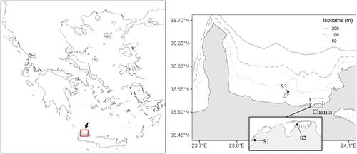
In the present study, we report the presence of different types of skeletal abnormalities in specimens of 10 fish species from the Aegean Sea. Estimations of abnormality prevalence are provided for seven species, whereas for three species we provide only single abnormality records. Fish samples were collected in Chania area (Crete, South Aegean Sea, Greece, Figure 1), both from recreational and commercial small-scale fisheries. Fish were transferred to the laboratory and digital images of the left side of each individual were captured. All specimens were then stored at −20°C (Laboratory of Marine Biology, Biology Department, University of Crete). Fish radiography was performed by using a Mex + 60 x-ray generator (medical ECONET GmbH, Oberhausen, Germany, 40–44 kV, 1.0–1.2 mA s, 75 cm focal distance) and a Dura-line XD 14 wireless DR detector (AGFA, Mortsel, Belgium).
A total of nine types of skeletal abnormalities were detected on jaws, vertebral column, dorsal and caudal fins, at rates of 3.3%–28.6% (Table 1). Vertebral abnormalities consisted of lordosis (V-shape bending of the vertebral column), kyphosis (Λ-shape bending of the vertebral column) and kypholordosis (concurrent lordosis and kyphosis) (Figures 2 and 3). In the studied Boops boops (Linnaeus, 1758) specimens, vertebral abnormalities consisted exclusively of kypholordosis (4.2%–10.3%), without any deviation from the normal meristic counts of the vertebrae and their processes (24 vertebrae) (Figure 2a,b). A single specimen of Mullus surmuletus (Linnaeus, 1758) presented concurrently three lordotic areas along its vertebral column (Figure 2c). Axis abnormalities in Sparisoma cretense (Linnaeus, 1758) (3.7%, Table 1) consisted of kyphosis of the haemal vertebral area, which was accompanied by an abnormally straightened anteriormost part of the axis, an upward shift of the skull (stargazer) and depressions of the dorsal profile of the body (Figure 2d,d′,e). A similar upward shift of the skull and depressions of the dorsal profile were observed in Pterois miles (Bennett, 1828) with lordosis (4.0%, Table 1) of the causal peduncle (Figure 3a,b). In this specimen, lordosis was associated with a fusion of the pre-ural centra 2 and 3 (PU2, PU3) into one bearing two haemal processes (Figure 3a′,b′). Finally, a Serranus scriba (Bennett, 1828) specimen (3.3%, Table 1) was recorded to present lordosis of the prehaemal area, linked with an upward shift of the skull (stargazer) (Figure 3c,d). Of the vertebral abnormalities recorded in the present study, kypholordosis is probably the most frequently reported in the wild fish (e.g., Bengtsson et al., 1985; Bensaada et al., 2023; Jawad et al., 2017; Jawad & Ibrahim, 2018a; Mariasingarayan et al., 2022; Van Den Avyle et al., 1989). On the other hand, to our knowledge, this is the first record of severe lordosis of the caudal peduncle, and of the abnormally straightened anteriormost part of the axis in natural fish populations.
In the present study, recorded skull abnormalities consisted of the compression of the ethmoid area and upper jaws (pugheadedness), shortened lower jaw, missing premaxillaries and lateral misalignment of the upper and lower jaws (crossbite). The highest frequency of pugheadedness was observed in one Diplodus sargus (Linnaeus, 1758) sample (28.6%, Table 1, Figure 4a–c). Pugheadedness and shortened lower jaw were present in both the examined samples of Pagellus erythrinus (Linnaeus, 1758) (Figure 4d–g), with equal frequencies (1.8%–2.8% for each abnormality type, Table 1). In one P. erythrinus specimen, pugheadedness was accompanied by crossbite (Figure 4e,e′). Missing premaxillaries were observed in one kypholordotic specimen of B. boops (Figure 4h). Of the skull abnormalities recorded in the present study, pugheadedness is the most frequently reported in wild fish (Bueno et al., 2015; Jawad et al., 2017; Näslund & Jawad, 2022; Porta & Snow, 2019). To our knowledge, crossbite and shortened lower jaw are frequently reported to develop in reared (Boglione et al., 2013; Fragkoulis et al., 2018), but rarely in wild fish.
In the present study, recorded fin abnormalities mainly consisted of the partial-to-complete lack of the dorsal fin, and/or depressions of the dorsal profile of the body (saddleback syndrome) (four species, Table 1). In one specimen of Siganus rivulatus Forsskål & Niebuhr (1775), the depression of the dorsal profile of the body was not associated with missing rays, but with a kyphosis of the adjacent vertebrae and abnormally shaped neural processes (Figure 5a,a′). In the second abnormal specimen of S. rivulatus detected, as well as in one S. cretense specimen, a dorsal body depression was associated with abnormal adjacent pterygiophores and missing rays (Figure 5b,b′,c,c′). A single specimen of D. sargus presented an anterior dorsal depression of the body, which was anatomically associated with abnormal shape and position of the predorsal elements (Figure 5d,d″). Finally, a single specimen of Symphodus tinca (Linnaeus, 1758) and Solea solea (Linnaeus, 1758) presented an entire missing part of its dorsal body area (Figure 5e,e′) and an entirely missing caudal fin (Figure 5f,f′) respectively. The term “saddleback syndrome” (SBS) was initially introduced by Tave et al. (1983) to characterize the partial-to-complete absence of the dorsal fin in tilapia Sarotherodon aureus. Since its inception, SBS has been observed to manifest in various fish species under captive conditions (Boglione et al., 2003; Cobcroft & Battaglene, 2013; Fragkoulis et al., 2017; Koumoundouros et al., 2001) and in wild fish populations globally (e.g., in 10 species by Browder et al., 1993; in S. cretense by Koumoundouros, 2008; in Acanthopagrus australis by Diggles, 2013; Pollock, 2015; in 10 species by Jawad & Ibrahim, 2018b; in Parastromateus niger by Silambarasan et al., 2021; in Lates calcarifer by Abed et al., 2023). Notably, Diggles (2013) documented an approximately sixfold increase in the prevalence of SBS among wild A. australis over a span of 20 years. The missing part of dorsal profile in S. tinca (present study) is similar to the abnormality reported in P. miles, which was attributed to physical injuries related to failed spearfishing attempts (Jimenez et al., 2022).
Skeletal abnormalities in fish may be induced by unfavourable environmental and nutritional conditions (reviewed by Boglione et al., 2013), genetic factors (e.g., Fragkoulis et al., 2020; Takeuchi, 1966; Tave et al., 1983), diseases (Ngo et al., 2024; Pasnik et al., 2007; Piamsomboon et al., 2022), pollutants (e.g., Johnson et al., 2020; Kessabi, Annabi, et al., 2013; Kessabi, Said, & Messaoudi, 2013; Messaoudi, Deli, et al., 2009), or toxins from harmful algal blooms (Sergi et al., 2022). In the present study, differences in abnormality rates and types among the studied species might be related to differences in life mode, feeding habits, reproduction pattern and seasonality. For example, B. boops juveniles migrate to the anthropogenically modified littoral zone (e.g., small harbours) (Georgiadis et al., 2014), where they may be subjected to various environmental stressors. A substantial number of abnormal specimens was collected by the present study in two small marinas. The high abnormality rates observed in the case of kypholordosis in B. boops (10.3%) and pugheadedness in D. sargus (28.6%) (Table 1), might be the result of localized causative factors, but also of the aggregation of deformed individuals in a protected environment where predation pressure is lower than in the open sea.
Publications reporting the presence of skeletal abnormalities in wild fish have been increasing in recent years. Since several of these abnormalities have significant adverse effects on fish performance (e.g., swimming, feeding), they could be considered as an additional source of mortality for natural stocks. However, the current available information does not allow for an estimation of the magnitude of the problem. This is mainly due to the lack of relevant monitoring surveys conducted at both spatial and temporal levels, which should also involve sampling at different developmental periods.
Dimitrios Chatzakis: Investigation; formal analysis. Roula Al Belbeisi: Investigation; formal analysis. Soultana Karagianni: Investigation; formal analysis. Eustratios Karagiannakidis: Visualization; methodology. George Koumoundouros: Formal analysis; writing – review and editing; writing – original draft; conceptualization; supervision.
This study was funded by the Property Development and Management Company of the University of Crete (Project no: 98).
期刊介绍:
Journal of Fish Diseases enjoys an international reputation as the medium for the exchange of information on original research into all aspects of disease in both wild and cultured fish and shellfish. Areas of interest regularly covered by the journal include:
-host-pathogen relationships-
studies of fish pathogens-
pathophysiology-
diagnostic methods-
therapy-
epidemiology-
descriptions of new diseases
 求助内容:
求助内容: 应助结果提醒方式:


