{"title":"利用全脑直方图和 Top20% 地图对淀粉样蛋白 PET/CT 进行客观简单评估的新方法。","authors":"Chio Okuyama, Tatsuya Higashi, Koichi Ishizu, Naoya Oishi, Kuninori Kusano, Miki Ito, Shinya Kagawa, Tomoko Okina, Norio Suzuki, Hiroshi Hasegawa, Yasuhiro Nagahama, Hiroyuki Watanabe, Masahiro Ono, Hiroshi Yamauchi","doi":"10.1007/s12149-024-01956-y","DOIUrl":null,"url":null,"abstract":"<div><h3>Objective</h3><p>This study aims to assess the utility of newly developed objective methods for the evaluation of intracranial abnormal amyloid deposition using PET/CT histogram without use of cortical ROI analyses.</p><h3>Methods</h3><p>Twenty-five healthy volunteers (HV) and 38 patients with diagnosed or suspected dementia who had undergone <sup>18</sup>F-FPYBF-2 PET/CT were retrospectively included in this study. Out of them, <sup>11</sup>C-PiB PET/CT had been also performed in 13 subjects. In addition to the conventional methods, namely visual judgment and quantitative analyses using composed standardized uptake value ratio (comSUVR), the PET images were also evaluated by the following new parameters: the skewness and the mode-to-mean ratio (MMR) obtained from the histogram of the brain parenchyma; Top20%-map highlights the areas with high tracer accumulation occupying 20% volume of the total brain parenchymal on the individual’s CT images. We evaluated the utility of the new methods using histogram compared with the visual assessment and comSUVR. The results of these new methods between <sup>18</sup>F-FPYBF-2 and <sup>11</sup>C-PiB were also compared in 13 subjects.</p><h3>Results</h3><p>In visual analysis, 32, 9, and 22 subjects showed negative, border, and positive results, and composed SUVR in each group were 1.11 ± 0.06, 1.20 ± 0.13, and 1.48 ± 0.18 (<i>p</i> < 0.0001), respectively. Visually positive subjects showed significantly low skewness and high MMR (<i>p</i> < 0.0001), and the Top20%-Map showed the presence or absence of abnormal deposits clearly. In comparison between the two tracers, visual evaluation was all consistent, and the ComSUVR, the skewness, the MMR showed significant good correlation. The Top20%-Maps showed similar pattern.</p><h3>Conclusions</h3><p>Our new methods using the histogram of the brain parenchymal accumulation are simple and suitable for clinical practice of amyloid PET, and Top20%-Map on the individual’s brain CT can be of great help for the visual assessment.</p></div>","PeriodicalId":8007,"journal":{"name":"Annals of Nuclear Medicine","volume":"38 9","pages":"763 - 773"},"PeriodicalIF":2.5000,"publicationDate":"2024-06-22","publicationTypes":"Journal Article","fieldsOfStudy":null,"isOpenAccess":false,"openAccessPdf":"https://www.ncbi.nlm.nih.gov/pmc/articles/PMC11339116/pdf/","citationCount":"0","resultStr":"{\"title\":\"New objective simple evaluation methods of amyloid PET/CT using whole-brain histogram and Top20%-Map\",\"authors\":\"Chio Okuyama, Tatsuya Higashi, Koichi Ishizu, Naoya Oishi, Kuninori Kusano, Miki Ito, Shinya Kagawa, Tomoko Okina, Norio Suzuki, Hiroshi Hasegawa, Yasuhiro Nagahama, Hiroyuki Watanabe, Masahiro Ono, Hiroshi Yamauchi\",\"doi\":\"10.1007/s12149-024-01956-y\",\"DOIUrl\":null,\"url\":null,\"abstract\":\"<div><h3>Objective</h3><p>This study aims to assess the utility of newly developed objective methods for the evaluation of intracranial abnormal amyloid deposition using PET/CT histogram without use of cortical ROI analyses.</p><h3>Methods</h3><p>Twenty-five healthy volunteers (HV) and 38 patients with diagnosed or suspected dementia who had undergone <sup>18</sup>F-FPYBF-2 PET/CT were retrospectively included in this study. Out of them, <sup>11</sup>C-PiB PET/CT had been also performed in 13 subjects. In addition to the conventional methods, namely visual judgment and quantitative analyses using composed standardized uptake value ratio (comSUVR), the PET images were also evaluated by the following new parameters: the skewness and the mode-to-mean ratio (MMR) obtained from the histogram of the brain parenchyma; Top20%-map highlights the areas with high tracer accumulation occupying 20% volume of the total brain parenchymal on the individual’s CT images. We evaluated the utility of the new methods using histogram compared with the visual assessment and comSUVR. The results of these new methods between <sup>18</sup>F-FPYBF-2 and <sup>11</sup>C-PiB were also compared in 13 subjects.</p><h3>Results</h3><p>In visual analysis, 32, 9, and 22 subjects showed negative, border, and positive results, and composed SUVR in each group were 1.11 ± 0.06, 1.20 ± 0.13, and 1.48 ± 0.18 (<i>p</i> < 0.0001), respectively. Visually positive subjects showed significantly low skewness and high MMR (<i>p</i> < 0.0001), and the Top20%-Map showed the presence or absence of abnormal deposits clearly. In comparison between the two tracers, visual evaluation was all consistent, and the ComSUVR, the skewness, the MMR showed significant good correlation. The Top20%-Maps showed similar pattern.</p><h3>Conclusions</h3><p>Our new methods using the histogram of the brain parenchymal accumulation are simple and suitable for clinical practice of amyloid PET, and Top20%-Map on the individual’s brain CT can be of great help for the visual assessment.</p></div>\",\"PeriodicalId\":8007,\"journal\":{\"name\":\"Annals of Nuclear Medicine\",\"volume\":\"38 9\",\"pages\":\"763 - 773\"},\"PeriodicalIF\":2.5000,\"publicationDate\":\"2024-06-22\",\"publicationTypes\":\"Journal Article\",\"fieldsOfStudy\":null,\"isOpenAccess\":false,\"openAccessPdf\":\"https://www.ncbi.nlm.nih.gov/pmc/articles/PMC11339116/pdf/\",\"citationCount\":\"0\",\"resultStr\":null,\"platform\":\"Semanticscholar\",\"paperid\":null,\"PeriodicalName\":\"Annals of Nuclear Medicine\",\"FirstCategoryId\":\"3\",\"ListUrlMain\":\"https://link.springer.com/article/10.1007/s12149-024-01956-y\",\"RegionNum\":4,\"RegionCategory\":\"医学\",\"ArticlePicture\":[],\"TitleCN\":null,\"AbstractTextCN\":null,\"PMCID\":null,\"EPubDate\":\"\",\"PubModel\":\"\",\"JCR\":\"Q2\",\"JCRName\":\"RADIOLOGY, NUCLEAR MEDICINE & MEDICAL IMAGING\",\"Score\":null,\"Total\":0}","platform":"Semanticscholar","paperid":null,"PeriodicalName":"Annals of Nuclear Medicine","FirstCategoryId":"3","ListUrlMain":"https://link.springer.com/article/10.1007/s12149-024-01956-y","RegionNum":4,"RegionCategory":"医学","ArticlePicture":[],"TitleCN":null,"AbstractTextCN":null,"PMCID":null,"EPubDate":"","PubModel":"","JCR":"Q2","JCRName":"RADIOLOGY, NUCLEAR MEDICINE & MEDICAL IMAGING","Score":null,"Total":0}
New objective simple evaluation methods of amyloid PET/CT using whole-brain histogram and Top20%-Map
Objective
This study aims to assess the utility of newly developed objective methods for the evaluation of intracranial abnormal amyloid deposition using PET/CT histogram without use of cortical ROI analyses.
Methods
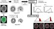
Twenty-five healthy volunteers (HV) and 38 patients with diagnosed or suspected dementia who had undergone 18F-FPYBF-2 PET/CT were retrospectively included in this study. Out of them, 11C-PiB PET/CT had been also performed in 13 subjects. In addition to the conventional methods, namely visual judgment and quantitative analyses using composed standardized uptake value ratio (comSUVR), the PET images were also evaluated by the following new parameters: the skewness and the mode-to-mean ratio (MMR) obtained from the histogram of the brain parenchyma; Top20%-map highlights the areas with high tracer accumulation occupying 20% volume of the total brain parenchymal on the individual’s CT images. We evaluated the utility of the new methods using histogram compared with the visual assessment and comSUVR. The results of these new methods between 18F-FPYBF-2 and 11C-PiB were also compared in 13 subjects.
Results
In visual analysis, 32, 9, and 22 subjects showed negative, border, and positive results, and composed SUVR in each group were 1.11 ± 0.06, 1.20 ± 0.13, and 1.48 ± 0.18 (p < 0.0001), respectively. Visually positive subjects showed significantly low skewness and high MMR (p < 0.0001), and the Top20%-Map showed the presence or absence of abnormal deposits clearly. In comparison between the two tracers, visual evaluation was all consistent, and the ComSUVR, the skewness, the MMR showed significant good correlation. The Top20%-Maps showed similar pattern.
Conclusions
Our new methods using the histogram of the brain parenchymal accumulation are simple and suitable for clinical practice of amyloid PET, and Top20%-Map on the individual’s brain CT can be of great help for the visual assessment.
期刊介绍:
Annals of Nuclear Medicine is an official journal of the Japanese Society of Nuclear Medicine. It develops the appropriate application of radioactive substances and stable nuclides in the field of medicine.
The journal promotes the exchange of ideas and information and research in nuclear medicine and includes the medical application of radionuclides and related subjects. It presents original articles, short communications, reviews and letters to the editor.

 求助内容:
求助内容: 应助结果提醒方式:


