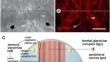
{"title":"额感觉-腺复合体及其神经投射的超微结构分析和三维重建。","authors":"Maria Del Mar de Miguel Bonet, Volker Hartenstein","doi":"10.1007/s00441-024-03901-x","DOIUrl":null,"url":null,"abstract":"<p><p>The marine microturbellarian Macrostomum lignano (Platyhelminthes, Rhabditophora) is an emerging laboratory model used by a growing community of researchers because it is easy to cultivate, has a fully sequenced genome, and offers multiple molecular tools for its study. M. lignano has a compartmentalized brain that receives sensory information from receptors integrated in the epidermis. Receptors of the head, as well as accompanying glands and specialized epidermal cells, form a compound sensory structure called the frontal glandular complex. In this study, we used semi-serial transmission electron microscopy (TEM) to document the types, ultrastructure, and three-dimensional architecture of the cells of the frontal glandular complex. We distinguish a ventral compartment formed by clusters of type 1 (multiciliated) sensory receptors from a central domain where type 2 (collar) sensory receptors predominate. Six different types of glands (rhammite glands, mucoid glands, glands with aster-like and perimaculate granula, vacuolated glands, and buckle glands) are closely associated with type 1 sensory receptors. Endings of a seventh type of gland (rhabdite gland) define a dorsal domain of the frontal glandular complex. A pair of ciliary photoreceptors is closely associated with the base of the frontal glandular complex. Bundles of dendrites, connecting the receptor endings with their cell bodies which are located in the brain, form the (frontal) peripheral nerves. Nerve fibers show a varicose structure, with thick segments alternating with thin segments, and are devoid of a glial layer. This distinguishes platyhelminths from larger and/or more complex invertebrates whose nerves are embedded in prominent glial sheaths.</p>","PeriodicalId":9712,"journal":{"name":"Cell and Tissue Research","volume":" ","pages":"147-177"},"PeriodicalIF":2.9000,"publicationDate":"2024-08-01","publicationTypes":"Journal Article","fieldsOfStudy":null,"isOpenAccess":false,"openAccessPdf":"","citationCount":"0","resultStr":"{\"title\":\"Ultrastructural analysis and 3D reconstruction of the frontal sensory-glandular complex and its neural projections in the platyhelminth Macrostomum lignano.\",\"authors\":\"Maria Del Mar de Miguel Bonet, Volker Hartenstein\",\"doi\":\"10.1007/s00441-024-03901-x\",\"DOIUrl\":null,\"url\":null,\"abstract\":\"<p><p>The marine microturbellarian Macrostomum lignano (Platyhelminthes, Rhabditophora) is an emerging laboratory model used by a growing community of researchers because it is easy to cultivate, has a fully sequenced genome, and offers multiple molecular tools for its study. M. lignano has a compartmentalized brain that receives sensory information from receptors integrated in the epidermis. Receptors of the head, as well as accompanying glands and specialized epidermal cells, form a compound sensory structure called the frontal glandular complex. In this study, we used semi-serial transmission electron microscopy (TEM) to document the types, ultrastructure, and three-dimensional architecture of the cells of the frontal glandular complex. We distinguish a ventral compartment formed by clusters of type 1 (multiciliated) sensory receptors from a central domain where type 2 (collar) sensory receptors predominate. Six different types of glands (rhammite glands, mucoid glands, glands with aster-like and perimaculate granula, vacuolated glands, and buckle glands) are closely associated with type 1 sensory receptors. Endings of a seventh type of gland (rhabdite gland) define a dorsal domain of the frontal glandular complex. A pair of ciliary photoreceptors is closely associated with the base of the frontal glandular complex. Bundles of dendrites, connecting the receptor endings with their cell bodies which are located in the brain, form the (frontal) peripheral nerves. Nerve fibers show a varicose structure, with thick segments alternating with thin segments, and are devoid of a glial layer. This distinguishes platyhelminths from larger and/or more complex invertebrates whose nerves are embedded in prominent glial sheaths.</p>\",\"PeriodicalId\":9712,\"journal\":{\"name\":\"Cell and Tissue Research\",\"volume\":\" \",\"pages\":\"147-177\"},\"PeriodicalIF\":2.9000,\"publicationDate\":\"2024-08-01\",\"publicationTypes\":\"Journal Article\",\"fieldsOfStudy\":null,\"isOpenAccess\":false,\"openAccessPdf\":\"\",\"citationCount\":\"0\",\"resultStr\":null,\"platform\":\"Semanticscholar\",\"paperid\":null,\"PeriodicalName\":\"Cell and Tissue Research\",\"FirstCategoryId\":\"99\",\"ListUrlMain\":\"https://doi.org/10.1007/s00441-024-03901-x\",\"RegionNum\":3,\"RegionCategory\":\"生物学\",\"ArticlePicture\":[],\"TitleCN\":null,\"AbstractTextCN\":null,\"PMCID\":null,\"EPubDate\":\"2024/6/20 0:00:00\",\"PubModel\":\"Epub\",\"JCR\":\"Q3\",\"JCRName\":\"CELL BIOLOGY\",\"Score\":null,\"Total\":0}","platform":"Semanticscholar","paperid":null,"PeriodicalName":"Cell and Tissue Research","FirstCategoryId":"99","ListUrlMain":"https://doi.org/10.1007/s00441-024-03901-x","RegionNum":3,"RegionCategory":"生物学","ArticlePicture":[],"TitleCN":null,"AbstractTextCN":null,"PMCID":null,"EPubDate":"2024/6/20 0:00:00","PubModel":"Epub","JCR":"Q3","JCRName":"CELL BIOLOGY","Score":null,"Total":0}
Ultrastructural analysis and 3D reconstruction of the frontal sensory-glandular complex and its neural projections in the platyhelminth Macrostomum lignano.
The marine microturbellarian Macrostomum lignano (Platyhelminthes, Rhabditophora) is an emerging laboratory model used by a growing community of researchers because it is easy to cultivate, has a fully sequenced genome, and offers multiple molecular tools for its study. M. lignano has a compartmentalized brain that receives sensory information from receptors integrated in the epidermis. Receptors of the head, as well as accompanying glands and specialized epidermal cells, form a compound sensory structure called the frontal glandular complex. In this study, we used semi-serial transmission electron microscopy (TEM) to document the types, ultrastructure, and three-dimensional architecture of the cells of the frontal glandular complex. We distinguish a ventral compartment formed by clusters of type 1 (multiciliated) sensory receptors from a central domain where type 2 (collar) sensory receptors predominate. Six different types of glands (rhammite glands, mucoid glands, glands with aster-like and perimaculate granula, vacuolated glands, and buckle glands) are closely associated with type 1 sensory receptors. Endings of a seventh type of gland (rhabdite gland) define a dorsal domain of the frontal glandular complex. A pair of ciliary photoreceptors is closely associated with the base of the frontal glandular complex. Bundles of dendrites, connecting the receptor endings with their cell bodies which are located in the brain, form the (frontal) peripheral nerves. Nerve fibers show a varicose structure, with thick segments alternating with thin segments, and are devoid of a glial layer. This distinguishes platyhelminths from larger and/or more complex invertebrates whose nerves are embedded in prominent glial sheaths.
期刊介绍:
The journal publishes regular articles and reviews in the areas of molecular, cell, and supracellular biology. In particular, the journal intends to provide a forum for publishing data that analyze the supracellular, integrative actions of gene products and their impact on the formation of tissue structure and function. Submission of papers with an emphasis on structure-function relationships as revealed by recombinant molecular technologies is especially encouraged. Areas of research with a long-standing tradition of publishing in Cell & Tissue Research include:
- neurobiology
- neuroendocrinology
- endocrinology
- reproductive biology
- skeletal and immune systems
- development
- stem cells
- muscle biology.
 求助内容:
求助内容: 应助结果提醒方式:


