Shah Ekramul Alom, Karthik Swaminathan, V Nuzelu, Alka Singh, Hugues de Rocquigny, Rajaram Swaminathan
{"title":"利用蛋白质电荷转移谱实时无标记跟踪乙型肝炎病毒核心蛋白囊壳组装。","authors":"Shah Ekramul Alom, Karthik Swaminathan, V Nuzelu, Alka Singh, Hugues de Rocquigny, Rajaram Swaminathan","doi":"10.1021/acs.biomac.4c00521","DOIUrl":null,"url":null,"abstract":"<p><p>Hepatitis B virions are double-shelled particles, with a diameter of 40-42 nm, consisting of a nucleocapsid called the HBV core protein (HBV Cp). It is an ordered assembly of 90-120 homodimers arranged in an icosahedral symmetry. Both the full-length HBV Cp and the first-149 residue domain, HBV Cp149, can spontaneously assemble in vitro into capsids with 120 Cp dimers (<i>T</i> = 4) or 90 Cp dimers (<i>T</i> = 3), triggered by high ionic strength of 0.25-0.5 M NaCl. The assembly disassembly of HBV Cp149 capsids are generally studied by light scattering, size-exclusion chromatography, atomic force microscopy, transmission electron microscopy, and other high-end expensive techniques. Here, we report a simple, yet robust, label-free technique exploiting protein charge transfer spectra (ProCharTS) to monitor the capsid assembly in real-time. ProCharTS absorption in the near UV-visible region (250-800 nm) arises when photoinduced electron transfer occurs from HOMO of COO<sup>-</sup> in glutamate (<i>donor</i>) to LUMO of NH<sub>3</sub><sup>+</sup> in lysine <i>or</i> polypeptide backbone (<i>acceptor</i>) of the protein. Alternatively, it can also occur from polypeptide backbone (<i>donor</i>) to <i>acceptor</i> in arginine, histidine, or lysine cation. ProCharTS is observed profusely among proximal charge clusters in folded proteins. Here, we show that, ProCharTS absorption among growing HBV capsids is amplified when HBV Cp homodimers assemble, generating new contacts among charged residues in the dimer-dimer interface. We notice a time-dependent sigmoidal increase in ProCharTS absorbance and luminescence during capsid formation in comparison to pure dimers. Additionally, a combined approach of anisotropy-based fluorescence assay is reported, where an increased fluorescence anisotropy was observed in capsids as compared to native and unfolded dimers. We conclude that ProCharTS can serve as a sensitive label-free tool for rapid tracking of capsid assembly in real-time and characterize the assembled capsids from dimers.</p>","PeriodicalId":30,"journal":{"name":"Biomacromolecules","volume":" ","pages":"6425-6438"},"PeriodicalIF":5.5000,"publicationDate":"2024-10-14","publicationTypes":"Journal Article","fieldsOfStudy":null,"isOpenAccess":false,"openAccessPdf":"","citationCount":"0","resultStr":"{\"title\":\"Label-Free Tracking of Hepatitis B Virus Core Protein Capsid Assembly in Real-Time Using Protein Charge Transfer Spectra.\",\"authors\":\"Shah Ekramul Alom, Karthik Swaminathan, V Nuzelu, Alka Singh, Hugues de Rocquigny, Rajaram Swaminathan\",\"doi\":\"10.1021/acs.biomac.4c00521\",\"DOIUrl\":null,\"url\":null,\"abstract\":\"<p><p>Hepatitis B virions are double-shelled particles, with a diameter of 40-42 nm, consisting of a nucleocapsid called the HBV core protein (HBV Cp). It is an ordered assembly of 90-120 homodimers arranged in an icosahedral symmetry. Both the full-length HBV Cp and the first-149 residue domain, HBV Cp149, can spontaneously assemble in vitro into capsids with 120 Cp dimers (<i>T</i> = 4) or 90 Cp dimers (<i>T</i> = 3), triggered by high ionic strength of 0.25-0.5 M NaCl. The assembly disassembly of HBV Cp149 capsids are generally studied by light scattering, size-exclusion chromatography, atomic force microscopy, transmission electron microscopy, and other high-end expensive techniques. Here, we report a simple, yet robust, label-free technique exploiting protein charge transfer spectra (ProCharTS) to monitor the capsid assembly in real-time. ProCharTS absorption in the near UV-visible region (250-800 nm) arises when photoinduced electron transfer occurs from HOMO of COO<sup>-</sup> in glutamate (<i>donor</i>) to LUMO of NH<sub>3</sub><sup>+</sup> in lysine <i>or</i> polypeptide backbone (<i>acceptor</i>) of the protein. Alternatively, it can also occur from polypeptide backbone (<i>donor</i>) to <i>acceptor</i> in arginine, histidine, or lysine cation. ProCharTS is observed profusely among proximal charge clusters in folded proteins. Here, we show that, ProCharTS absorption among growing HBV capsids is amplified when HBV Cp homodimers assemble, generating new contacts among charged residues in the dimer-dimer interface. We notice a time-dependent sigmoidal increase in ProCharTS absorbance and luminescence during capsid formation in comparison to pure dimers. Additionally, a combined approach of anisotropy-based fluorescence assay is reported, where an increased fluorescence anisotropy was observed in capsids as compared to native and unfolded dimers. We conclude that ProCharTS can serve as a sensitive label-free tool for rapid tracking of capsid assembly in real-time and characterize the assembled capsids from dimers.</p>\",\"PeriodicalId\":30,\"journal\":{\"name\":\"Biomacromolecules\",\"volume\":\" \",\"pages\":\"6425-6438\"},\"PeriodicalIF\":5.5000,\"publicationDate\":\"2024-10-14\",\"publicationTypes\":\"Journal Article\",\"fieldsOfStudy\":null,\"isOpenAccess\":false,\"openAccessPdf\":\"\",\"citationCount\":\"0\",\"resultStr\":null,\"platform\":\"Semanticscholar\",\"paperid\":null,\"PeriodicalName\":\"Biomacromolecules\",\"FirstCategoryId\":\"92\",\"ListUrlMain\":\"https://doi.org/10.1021/acs.biomac.4c00521\",\"RegionNum\":2,\"RegionCategory\":\"化学\",\"ArticlePicture\":[],\"TitleCN\":null,\"AbstractTextCN\":null,\"PMCID\":null,\"EPubDate\":\"2024/6/20 0:00:00\",\"PubModel\":\"Epub\",\"JCR\":\"Q1\",\"JCRName\":\"BIOCHEMISTRY & MOLECULAR BIOLOGY\",\"Score\":null,\"Total\":0}","platform":"Semanticscholar","paperid":null,"PeriodicalName":"Biomacromolecules","FirstCategoryId":"92","ListUrlMain":"https://doi.org/10.1021/acs.biomac.4c00521","RegionNum":2,"RegionCategory":"化学","ArticlePicture":[],"TitleCN":null,"AbstractTextCN":null,"PMCID":null,"EPubDate":"2024/6/20 0:00:00","PubModel":"Epub","JCR":"Q1","JCRName":"BIOCHEMISTRY & MOLECULAR BIOLOGY","Score":null,"Total":0}
Label-Free Tracking of Hepatitis B Virus Core Protein Capsid Assembly in Real-Time Using Protein Charge Transfer Spectra.
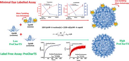
Hepatitis B virions are double-shelled particles, with a diameter of 40-42 nm, consisting of a nucleocapsid called the HBV core protein (HBV Cp). It is an ordered assembly of 90-120 homodimers arranged in an icosahedral symmetry. Both the full-length HBV Cp and the first-149 residue domain, HBV Cp149, can spontaneously assemble in vitro into capsids with 120 Cp dimers (T = 4) or 90 Cp dimers (T = 3), triggered by high ionic strength of 0.25-0.5 M NaCl. The assembly disassembly of HBV Cp149 capsids are generally studied by light scattering, size-exclusion chromatography, atomic force microscopy, transmission electron microscopy, and other high-end expensive techniques. Here, we report a simple, yet robust, label-free technique exploiting protein charge transfer spectra (ProCharTS) to monitor the capsid assembly in real-time. ProCharTS absorption in the near UV-visible region (250-800 nm) arises when photoinduced electron transfer occurs from HOMO of COO- in glutamate (donor) to LUMO of NH3+ in lysine or polypeptide backbone (acceptor) of the protein. Alternatively, it can also occur from polypeptide backbone (donor) to acceptor in arginine, histidine, or lysine cation. ProCharTS is observed profusely among proximal charge clusters in folded proteins. Here, we show that, ProCharTS absorption among growing HBV capsids is amplified when HBV Cp homodimers assemble, generating new contacts among charged residues in the dimer-dimer interface. We notice a time-dependent sigmoidal increase in ProCharTS absorbance and luminescence during capsid formation in comparison to pure dimers. Additionally, a combined approach of anisotropy-based fluorescence assay is reported, where an increased fluorescence anisotropy was observed in capsids as compared to native and unfolded dimers. We conclude that ProCharTS can serve as a sensitive label-free tool for rapid tracking of capsid assembly in real-time and characterize the assembled capsids from dimers.
期刊介绍:
Biomacromolecules is a leading forum for the dissemination of cutting-edge research at the interface of polymer science and biology. Submissions to Biomacromolecules should contain strong elements of innovation in terms of macromolecular design, synthesis and characterization, or in the application of polymer materials to biology and medicine.
Topics covered by Biomacromolecules include, but are not exclusively limited to: sustainable polymers, polymers based on natural and renewable resources, degradable polymers, polymer conjugates, polymeric drugs, polymers in biocatalysis, biomacromolecular assembly, biomimetic polymers, polymer-biomineral hybrids, biomimetic-polymer processing, polymer recycling, bioactive polymer surfaces, original polymer design for biomedical applications such as immunotherapy, drug delivery, gene delivery, antimicrobial applications, diagnostic imaging and biosensing, polymers in tissue engineering and regenerative medicine, polymeric scaffolds and hydrogels for cell culture and delivery.
 求助内容:
求助内容: 应助结果提醒方式:


