{"title":"通过计算机断层扫描图像上的不对称扩散来区分鼻腭管囊肿和上颌骨前部的根状囊肿。","authors":"Haruka Ikeda, Natsuho Takata, Yoshitaka Kise, Kaori Ebata, Mizuho Mori, Chiaki Kuwada, Masako Nishiyama, Yukiko Iwase, Yo Ninagawa, Munetaka Naitoh, Eiichiro Ariji","doi":"10.1007/s11282-024-00761-7","DOIUrl":null,"url":null,"abstract":"<p><strong>Objective: </strong>The aim of this study was to clarify numerical values for differentiating nasopalatine duct cysts (NPDCs) from radicular cysts (RCs) arising in the anterior maxilla on computed tomography (CT) or cone-beam CT (CBCT) images.</p><p><strong>Methods: </strong>CT or CBCT images of histologically proven NPDCs (n = 30) and RCs (n = 33) beyond the midline of the maxilla were investigated to determine two asymmetry indices on axial images of the maximum lesion area. The lateral asymmetry index was calculated based on two distances from each of the lateral ends of the lesion to the midsagittal plane. The index was defined as the difference between the two distances divided by their sum. The labio-palatal asymmetry index was determined by the distance between the labial and palatal ends of the lesion and the coronal plane passing through the central incisor root apex. The performance of these indices was assessed by receiver operating characteristic (ROC) analysis. The cutoff values for differentiating NPDCs from RCs were determined with the Youden procedure on the ROC curve.</p><p><strong>Results: </strong>The area under the ROC curve was 0.97 for the lateral asymmetry index and 0.88 for the labio-palatal asymmetry index. The cutoff values for differentiation were 0.36 and 0.68 for the lateral and labio-palatal asymmetry indices, respectively.</p><p><strong>Conclusion: </strong>The lateral asymmetry index appeared to be an effective reference for differentiating NPDCs from RCs on CT or CBCT images. When the index was less than the cutoff value, a diagnosis of NPDC was strongly suggested.</p>","PeriodicalId":56103,"journal":{"name":"Oral Radiology","volume":" ","pages":"501-507"},"PeriodicalIF":1.6000,"publicationDate":"2024-10-01","publicationTypes":"Journal Article","fieldsOfStudy":null,"isOpenAccess":false,"openAccessPdf":"","citationCount":"0","resultStr":"{\"title\":\"Spread asymmetry to differentiate nasopalatine duct cysts from radicular cysts arising in the anterior maxilla on computed tomographic images.\",\"authors\":\"Haruka Ikeda, Natsuho Takata, Yoshitaka Kise, Kaori Ebata, Mizuho Mori, Chiaki Kuwada, Masako Nishiyama, Yukiko Iwase, Yo Ninagawa, Munetaka Naitoh, Eiichiro Ariji\",\"doi\":\"10.1007/s11282-024-00761-7\",\"DOIUrl\":null,\"url\":null,\"abstract\":\"<p><strong>Objective: </strong>The aim of this study was to clarify numerical values for differentiating nasopalatine duct cysts (NPDCs) from radicular cysts (RCs) arising in the anterior maxilla on computed tomography (CT) or cone-beam CT (CBCT) images.</p><p><strong>Methods: </strong>CT or CBCT images of histologically proven NPDCs (n = 30) and RCs (n = 33) beyond the midline of the maxilla were investigated to determine two asymmetry indices on axial images of the maximum lesion area. The lateral asymmetry index was calculated based on two distances from each of the lateral ends of the lesion to the midsagittal plane. The index was defined as the difference between the two distances divided by their sum. The labio-palatal asymmetry index was determined by the distance between the labial and palatal ends of the lesion and the coronal plane passing through the central incisor root apex. The performance of these indices was assessed by receiver operating characteristic (ROC) analysis. The cutoff values for differentiating NPDCs from RCs were determined with the Youden procedure on the ROC curve.</p><p><strong>Results: </strong>The area under the ROC curve was 0.97 for the lateral asymmetry index and 0.88 for the labio-palatal asymmetry index. The cutoff values for differentiation were 0.36 and 0.68 for the lateral and labio-palatal asymmetry indices, respectively.</p><p><strong>Conclusion: </strong>The lateral asymmetry index appeared to be an effective reference for differentiating NPDCs from RCs on CT or CBCT images. When the index was less than the cutoff value, a diagnosis of NPDC was strongly suggested.</p>\",\"PeriodicalId\":56103,\"journal\":{\"name\":\"Oral Radiology\",\"volume\":\" \",\"pages\":\"501-507\"},\"PeriodicalIF\":1.6000,\"publicationDate\":\"2024-10-01\",\"publicationTypes\":\"Journal Article\",\"fieldsOfStudy\":null,\"isOpenAccess\":false,\"openAccessPdf\":\"\",\"citationCount\":\"0\",\"resultStr\":null,\"platform\":\"Semanticscholar\",\"paperid\":null,\"PeriodicalName\":\"Oral Radiology\",\"FirstCategoryId\":\"3\",\"ListUrlMain\":\"https://doi.org/10.1007/s11282-024-00761-7\",\"RegionNum\":3,\"RegionCategory\":\"医学\",\"ArticlePicture\":[],\"TitleCN\":null,\"AbstractTextCN\":null,\"PMCID\":null,\"EPubDate\":\"2024/6/18 0:00:00\",\"PubModel\":\"Epub\",\"JCR\":\"Q3\",\"JCRName\":\"DENTISTRY, ORAL SURGERY & MEDICINE\",\"Score\":null,\"Total\":0}","platform":"Semanticscholar","paperid":null,"PeriodicalName":"Oral Radiology","FirstCategoryId":"3","ListUrlMain":"https://doi.org/10.1007/s11282-024-00761-7","RegionNum":3,"RegionCategory":"医学","ArticlePicture":[],"TitleCN":null,"AbstractTextCN":null,"PMCID":null,"EPubDate":"2024/6/18 0:00:00","PubModel":"Epub","JCR":"Q3","JCRName":"DENTISTRY, ORAL SURGERY & MEDICINE","Score":null,"Total":0}
Spread asymmetry to differentiate nasopalatine duct cysts from radicular cysts arising in the anterior maxilla on computed tomographic images.
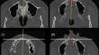
Objective: The aim of this study was to clarify numerical values for differentiating nasopalatine duct cysts (NPDCs) from radicular cysts (RCs) arising in the anterior maxilla on computed tomography (CT) or cone-beam CT (CBCT) images.
Methods: CT or CBCT images of histologically proven NPDCs (n = 30) and RCs (n = 33) beyond the midline of the maxilla were investigated to determine two asymmetry indices on axial images of the maximum lesion area. The lateral asymmetry index was calculated based on two distances from each of the lateral ends of the lesion to the midsagittal plane. The index was defined as the difference between the two distances divided by their sum. The labio-palatal asymmetry index was determined by the distance between the labial and palatal ends of the lesion and the coronal plane passing through the central incisor root apex. The performance of these indices was assessed by receiver operating characteristic (ROC) analysis. The cutoff values for differentiating NPDCs from RCs were determined with the Youden procedure on the ROC curve.
Results: The area under the ROC curve was 0.97 for the lateral asymmetry index and 0.88 for the labio-palatal asymmetry index. The cutoff values for differentiation were 0.36 and 0.68 for the lateral and labio-palatal asymmetry indices, respectively.
Conclusion: The lateral asymmetry index appeared to be an effective reference for differentiating NPDCs from RCs on CT or CBCT images. When the index was less than the cutoff value, a diagnosis of NPDC was strongly suggested.
期刊介绍:
As the official English-language journal of the Japanese Society for Oral and Maxillofacial Radiology and the Asian Academy of Oral and Maxillofacial Radiology, Oral Radiology is intended to be a forum for international collaboration in head and neck diagnostic imaging and all related fields. Oral Radiology features cutting-edge research papers, review articles, case reports, and technical notes from both the clinical and experimental fields. As membership in the Society is not a prerequisite, contributions are welcome from researchers and clinicians worldwide.

 求助内容:
求助内容: 应助结果提醒方式:


