{"title":"结肠活检中免疫检查点抑制剂治疗后未掩盖的炎症性肠病的组织病理学特征","authors":"","doi":"10.1016/j.gastha.2024.05.011","DOIUrl":null,"url":null,"abstract":"<div><h3>Background and Aims</h3><p>Typical immune checkpoint inhibitor-induced colitis (T-ICI) has significant histomorphologic overlap with inflammatory bowel disease (IBD), a distinction further complicated in ICI-treated patients with pre-existing inflammatory bowel disease (P-IBD) and those with potentially “unmasked” inflammatory bowel disease (U-IBD) after ICI therapy. This study describes histopathologic findings seen in U-IBD colonic biopsies and assesses for distinguishing features from T-ICI and P-IBD biopsies.</p></div><div><h3>Methods</h3><p>Initial colon biopsies after symptom onset from 34 patients on ICI therapy were reviewed, and histopathologic features were tabulated. U-IBD patients were identified clinically based on rapid toxicity development post-ICI treatment with multiple recurrences after immune suppression, frequently with regional colitis (versus pancolitis).</p></div><div><h3>Results</h3><p>The study cohort was classified into T-ICI (n = 20), P-IBD (n = 9), and U-IBD (n = 5) groups. The predominant histological patterns were diffuse active colitis (35%) in the T-ICI, and chronic active colitis in both the P-IBD (67%) and U-IBD (60%) groups (overall <em>P</em> = .003, <em>P</em> > .05 between the two IBD groups). None of the T-ICI biopsies demonstrated chronicity features (ie, architectural distortion score 2, basal lymphoplasmacytosis, or Paneth cell metaplasia). Only U-IBD biopsies demonstrated basal lymphoplasmacytosis (60% vs 0% in T-ICI/P-IBD, <em>P</em> = .002). Among available follow-up biopsies, chronicity features were present in all (4/4) U-IBD patients, including those without chronicity seen in the initial biopsy, but none (0/7) of T-ICI patients.</p></div><div><h3>Conclusion</h3><p>These early results show that no definite features of chronicity were seen in colon biopsies from T-ICI patients, suggesting that the presence of those features may be a clue to U-IBD in patients without a known IBD diagnosis. Frequent basal lymphoplasmacytosis seen in U-IBD may support a recent onset of mucosal injury and early architectural remodeling.</p></div>","PeriodicalId":73130,"journal":{"name":"Gastro hep advances","volume":"3 7","pages":"Pages 986-994"},"PeriodicalIF":0.0000,"publicationDate":"2024-01-01","publicationTypes":"Journal Article","fieldsOfStudy":null,"isOpenAccess":false,"openAccessPdf":"https://www.sciencedirect.com/science/article/pii/S2772572324000797/pdfft?md5=ddf2747df63531821784e978adf831b8&pid=1-s2.0-S2772572324000797-main.pdf","citationCount":"0","resultStr":"{\"title\":\"Histopathologic Features of Unmasked Inflammatory Bowel Disease Following Immune Checkpoint Inhibitor Therapy in Colon Biopsies\",\"authors\":\"\",\"doi\":\"10.1016/j.gastha.2024.05.011\",\"DOIUrl\":null,\"url\":null,\"abstract\":\"<div><h3>Background and Aims</h3><p>Typical immune checkpoint inhibitor-induced colitis (T-ICI) has significant histomorphologic overlap with inflammatory bowel disease (IBD), a distinction further complicated in ICI-treated patients with pre-existing inflammatory bowel disease (P-IBD) and those with potentially “unmasked” inflammatory bowel disease (U-IBD) after ICI therapy. This study describes histopathologic findings seen in U-IBD colonic biopsies and assesses for distinguishing features from T-ICI and P-IBD biopsies.</p></div><div><h3>Methods</h3><p>Initial colon biopsies after symptom onset from 34 patients on ICI therapy were reviewed, and histopathologic features were tabulated. U-IBD patients were identified clinically based on rapid toxicity development post-ICI treatment with multiple recurrences after immune suppression, frequently with regional colitis (versus pancolitis).</p></div><div><h3>Results</h3><p>The study cohort was classified into T-ICI (n = 20), P-IBD (n = 9), and U-IBD (n = 5) groups. The predominant histological patterns were diffuse active colitis (35%) in the T-ICI, and chronic active colitis in both the P-IBD (67%) and U-IBD (60%) groups (overall <em>P</em> = .003, <em>P</em> > .05 between the two IBD groups). None of the T-ICI biopsies demonstrated chronicity features (ie, architectural distortion score 2, basal lymphoplasmacytosis, or Paneth cell metaplasia). Only U-IBD biopsies demonstrated basal lymphoplasmacytosis (60% vs 0% in T-ICI/P-IBD, <em>P</em> = .002). Among available follow-up biopsies, chronicity features were present in all (4/4) U-IBD patients, including those without chronicity seen in the initial biopsy, but none (0/7) of T-ICI patients.</p></div><div><h3>Conclusion</h3><p>These early results show that no definite features of chronicity were seen in colon biopsies from T-ICI patients, suggesting that the presence of those features may be a clue to U-IBD in patients without a known IBD diagnosis. Frequent basal lymphoplasmacytosis seen in U-IBD may support a recent onset of mucosal injury and early architectural remodeling.</p></div>\",\"PeriodicalId\":73130,\"journal\":{\"name\":\"Gastro hep advances\",\"volume\":\"3 7\",\"pages\":\"Pages 986-994\"},\"PeriodicalIF\":0.0000,\"publicationDate\":\"2024-01-01\",\"publicationTypes\":\"Journal Article\",\"fieldsOfStudy\":null,\"isOpenAccess\":false,\"openAccessPdf\":\"https://www.sciencedirect.com/science/article/pii/S2772572324000797/pdfft?md5=ddf2747df63531821784e978adf831b8&pid=1-s2.0-S2772572324000797-main.pdf\",\"citationCount\":\"0\",\"resultStr\":null,\"platform\":\"Semanticscholar\",\"paperid\":null,\"PeriodicalName\":\"Gastro hep advances\",\"FirstCategoryId\":\"1085\",\"ListUrlMain\":\"https://www.sciencedirect.com/science/article/pii/S2772572324000797\",\"RegionNum\":0,\"RegionCategory\":null,\"ArticlePicture\":[],\"TitleCN\":null,\"AbstractTextCN\":null,\"PMCID\":null,\"EPubDate\":\"\",\"PubModel\":\"\",\"JCR\":\"\",\"JCRName\":\"\",\"Score\":null,\"Total\":0}","platform":"Semanticscholar","paperid":null,"PeriodicalName":"Gastro hep advances","FirstCategoryId":"1085","ListUrlMain":"https://www.sciencedirect.com/science/article/pii/S2772572324000797","RegionNum":0,"RegionCategory":null,"ArticlePicture":[],"TitleCN":null,"AbstractTextCN":null,"PMCID":null,"EPubDate":"","PubModel":"","JCR":"","JCRName":"","Score":null,"Total":0}
Histopathologic Features of Unmasked Inflammatory Bowel Disease Following Immune Checkpoint Inhibitor Therapy in Colon Biopsies
Background and Aims
Typical immune checkpoint inhibitor-induced colitis (T-ICI) has significant histomorphologic overlap with inflammatory bowel disease (IBD), a distinction further complicated in ICI-treated patients with pre-existing inflammatory bowel disease (P-IBD) and those with potentially “unmasked” inflammatory bowel disease (U-IBD) after ICI therapy. This study describes histopathologic findings seen in U-IBD colonic biopsies and assesses for distinguishing features from T-ICI and P-IBD biopsies.
Methods
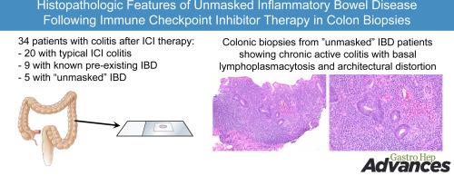
Initial colon biopsies after symptom onset from 34 patients on ICI therapy were reviewed, and histopathologic features were tabulated. U-IBD patients were identified clinically based on rapid toxicity development post-ICI treatment with multiple recurrences after immune suppression, frequently with regional colitis (versus pancolitis).
Results
The study cohort was classified into T-ICI (n = 20), P-IBD (n = 9), and U-IBD (n = 5) groups. The predominant histological patterns were diffuse active colitis (35%) in the T-ICI, and chronic active colitis in both the P-IBD (67%) and U-IBD (60%) groups (overall P = .003, P > .05 between the two IBD groups). None of the T-ICI biopsies demonstrated chronicity features (ie, architectural distortion score 2, basal lymphoplasmacytosis, or Paneth cell metaplasia). Only U-IBD biopsies demonstrated basal lymphoplasmacytosis (60% vs 0% in T-ICI/P-IBD, P = .002). Among available follow-up biopsies, chronicity features were present in all (4/4) U-IBD patients, including those without chronicity seen in the initial biopsy, but none (0/7) of T-ICI patients.
Conclusion
These early results show that no definite features of chronicity were seen in colon biopsies from T-ICI patients, suggesting that the presence of those features may be a clue to U-IBD in patients without a known IBD diagnosis. Frequent basal lymphoplasmacytosis seen in U-IBD may support a recent onset of mucosal injury and early architectural remodeling.

 求助内容:
求助内容: 应助结果提醒方式:


