Tiziano Balzano, Natalia López-González del Rey, Noelia Esteban-García, Alejandro Reinares-Sebastián, José A. Pineda-Pardo, Inés Trigo-Damas, José A. Obeso, Javier Blesa
{"title":"非人灵长类黑质多巴胺能神经元易损性的神经血管和免疫因素","authors":"Tiziano Balzano, Natalia López-González del Rey, Noelia Esteban-García, Alejandro Reinares-Sebastián, José A. Pineda-Pardo, Inés Trigo-Damas, José A. Obeso, Javier Blesa","doi":"10.1038/s41531-024-00735-w","DOIUrl":null,"url":null,"abstract":"<p>Dopaminergic neurons in the ventral tier of the substantia nigra pars compacta (SNc) degenerate prominently in Parkinson’s disease (PD), while those in the dorsal tier and ventral tegmental area are relatively spared. The factors determining why these neurons are more vulnerable than others are still unrevealed. Neuroinflammation and immune cell infiltration have been demonstrated to be a key feature of neurodegeneration in PD. However, the link between selective dopaminergic neuron vulnerability, glial and immune cell response, and vascularization and their interactions has not been deciphered. We aimed to investigate the contribution of glial cell activation and immune cell infiltration in the selective vulnerability of ventral dopaminergic neurons within the midbrain in a non-human primate model of PD. Structural characteristics of the vasculature within specific regions of the midbrain were also evaluated. Parkinsonian monkeys exhibited significant microglial and astroglial activation in the whole midbrain, but no major sub-regional differences were observed. Remarkably, the ventral substantia nigra was found to be typically more vascularized compared to other regions. This feature might play some role in making this region more susceptible to immune cell infiltration under pathological conditions, as greater infiltration of both T- and B- lymphocytes was observed in parkinsonian monkeys. Higher vascular density within the ventral region of the SNc may be a relevant factor for differential vulnerability of dopaminergic neurons in the midbrain. The increased infiltration of T- and B- cells in this region, alongside other molecules or toxins, may also contribute to the susceptibility of dopaminergic neurons in PD.</p>","PeriodicalId":19706,"journal":{"name":"NPJ Parkinson's Disease","volume":null,"pages":null},"PeriodicalIF":6.7000,"publicationDate":"2024-06-17","publicationTypes":"Journal Article","fieldsOfStudy":null,"isOpenAccess":false,"openAccessPdf":"","citationCount":"0","resultStr":"{\"title\":\"Neurovascular and immune factors of vulnerability of substantia nigra dopaminergic neurons in non-human primates\",\"authors\":\"Tiziano Balzano, Natalia López-González del Rey, Noelia Esteban-García, Alejandro Reinares-Sebastián, José A. Pineda-Pardo, Inés Trigo-Damas, José A. Obeso, Javier Blesa\",\"doi\":\"10.1038/s41531-024-00735-w\",\"DOIUrl\":null,\"url\":null,\"abstract\":\"<p>Dopaminergic neurons in the ventral tier of the substantia nigra pars compacta (SNc) degenerate prominently in Parkinson’s disease (PD), while those in the dorsal tier and ventral tegmental area are relatively spared. The factors determining why these neurons are more vulnerable than others are still unrevealed. Neuroinflammation and immune cell infiltration have been demonstrated to be a key feature of neurodegeneration in PD. However, the link between selective dopaminergic neuron vulnerability, glial and immune cell response, and vascularization and their interactions has not been deciphered. We aimed to investigate the contribution of glial cell activation and immune cell infiltration in the selective vulnerability of ventral dopaminergic neurons within the midbrain in a non-human primate model of PD. Structural characteristics of the vasculature within specific regions of the midbrain were also evaluated. Parkinsonian monkeys exhibited significant microglial and astroglial activation in the whole midbrain, but no major sub-regional differences were observed. Remarkably, the ventral substantia nigra was found to be typically more vascularized compared to other regions. This feature might play some role in making this region more susceptible to immune cell infiltration under pathological conditions, as greater infiltration of both T- and B- lymphocytes was observed in parkinsonian monkeys. Higher vascular density within the ventral region of the SNc may be a relevant factor for differential vulnerability of dopaminergic neurons in the midbrain. The increased infiltration of T- and B- cells in this region, alongside other molecules or toxins, may also contribute to the susceptibility of dopaminergic neurons in PD.</p>\",\"PeriodicalId\":19706,\"journal\":{\"name\":\"NPJ Parkinson's Disease\",\"volume\":null,\"pages\":null},\"PeriodicalIF\":6.7000,\"publicationDate\":\"2024-06-17\",\"publicationTypes\":\"Journal Article\",\"fieldsOfStudy\":null,\"isOpenAccess\":false,\"openAccessPdf\":\"\",\"citationCount\":\"0\",\"resultStr\":null,\"platform\":\"Semanticscholar\",\"paperid\":null,\"PeriodicalName\":\"NPJ Parkinson's Disease\",\"FirstCategoryId\":\"3\",\"ListUrlMain\":\"https://doi.org/10.1038/s41531-024-00735-w\",\"RegionNum\":1,\"RegionCategory\":\"医学\",\"ArticlePicture\":[],\"TitleCN\":null,\"AbstractTextCN\":null,\"PMCID\":null,\"EPubDate\":\"\",\"PubModel\":\"\",\"JCR\":\"Q1\",\"JCRName\":\"NEUROSCIENCES\",\"Score\":null,\"Total\":0}","platform":"Semanticscholar","paperid":null,"PeriodicalName":"NPJ Parkinson's Disease","FirstCategoryId":"3","ListUrlMain":"https://doi.org/10.1038/s41531-024-00735-w","RegionNum":1,"RegionCategory":"医学","ArticlePicture":[],"TitleCN":null,"AbstractTextCN":null,"PMCID":null,"EPubDate":"","PubModel":"","JCR":"Q1","JCRName":"NEUROSCIENCES","Score":null,"Total":0}
引用次数: 0
摘要
帕金森病(Parkinson's disease,PD)患者黑质紧密团(substantia nigra pars compacta,SNc)腹侧层的多巴胺能神经元会显著退化,而背侧层和腹侧被盖区的多巴胺能神经元则相对幸免。决定这些神经元比其他神经元更脆弱的因素仍未揭示。神经炎症和免疫细胞浸润已被证实是帕金森病神经变性的一个主要特征。然而,选择性多巴胺能神经元的脆弱性、神经胶质细胞和免疫细胞的反应以及血管化之间的联系及其相互作用尚未被破解。我们旨在研究神经胶质细胞活化和免疫细胞浸润在非人灵长类疾病模型中中脑腹侧多巴胺能神经元选择性易损性中的作用。此外,还评估了中脑特定区域内血管的结构特征。帕金森病猴的整个中脑表现出明显的小胶质细胞和星形胶质细胞活化,但没有观察到重大的亚区域差异。值得注意的是,与其他区域相比,腹侧黑质的血管化程度通常更高。在帕金森病猴身上观察到更多的T淋巴细胞和B淋巴细胞浸润,这一特征可能在某种程度上使该区域在病理条件下更容易受到免疫细胞的浸润。SNc腹侧区域内较高的血管密度可能是导致中脑多巴胺能神经元不同脆弱性的一个相关因素。该区域T细胞和B细胞浸润的增加,以及其他分子或毒素,也可能导致帕金森病多巴胺能神经元的易感性。
Neurovascular and immune factors of vulnerability of substantia nigra dopaminergic neurons in non-human primates
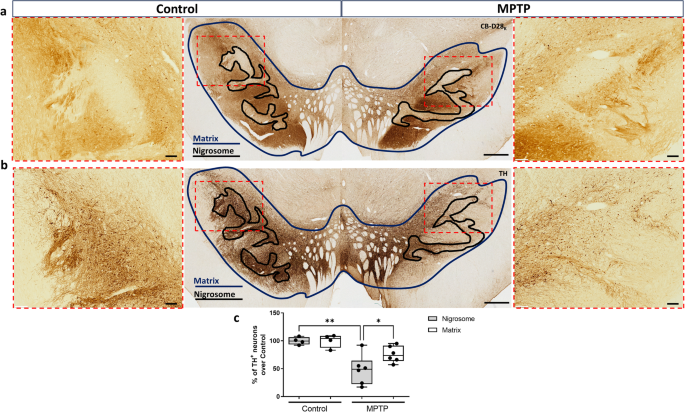
Dopaminergic neurons in the ventral tier of the substantia nigra pars compacta (SNc) degenerate prominently in Parkinson’s disease (PD), while those in the dorsal tier and ventral tegmental area are relatively spared. The factors determining why these neurons are more vulnerable than others are still unrevealed. Neuroinflammation and immune cell infiltration have been demonstrated to be a key feature of neurodegeneration in PD. However, the link between selective dopaminergic neuron vulnerability, glial and immune cell response, and vascularization and their interactions has not been deciphered. We aimed to investigate the contribution of glial cell activation and immune cell infiltration in the selective vulnerability of ventral dopaminergic neurons within the midbrain in a non-human primate model of PD. Structural characteristics of the vasculature within specific regions of the midbrain were also evaluated. Parkinsonian monkeys exhibited significant microglial and astroglial activation in the whole midbrain, but no major sub-regional differences were observed. Remarkably, the ventral substantia nigra was found to be typically more vascularized compared to other regions. This feature might play some role in making this region more susceptible to immune cell infiltration under pathological conditions, as greater infiltration of both T- and B- lymphocytes was observed in parkinsonian monkeys. Higher vascular density within the ventral region of the SNc may be a relevant factor for differential vulnerability of dopaminergic neurons in the midbrain. The increased infiltration of T- and B- cells in this region, alongside other molecules or toxins, may also contribute to the susceptibility of dopaminergic neurons in PD.
期刊介绍:
npj Parkinson's Disease is a comprehensive open access journal that covers a wide range of research areas related to Parkinson's disease. It publishes original studies in basic science, translational research, and clinical investigations. The journal is dedicated to advancing our understanding of Parkinson's disease by exploring various aspects such as anatomy, etiology, genetics, cellular and molecular physiology, neurophysiology, epidemiology, and therapeutic development. By providing free and immediate access to the scientific and Parkinson's disease community, npj Parkinson's Disease promotes collaboration and knowledge sharing among researchers and healthcare professionals.
 求助内容:
求助内容: 应助结果提醒方式:


