Caner Ercan, Kattayoun Kordy, Anna Knuuttila, Xiaofei Zhou, Darshan Kumar, Ville Koponen, Peter Mesenbrink, Serenella Eppenberger-Castori, Parisa Amini, Marcos C Pedrosa, Luigi M Terracciano
{"title":"基于深度学习的组织学自身免疫性肝炎评估模型:AI(H).","authors":"Caner Ercan, Kattayoun Kordy, Anna Knuuttila, Xiaofei Zhou, Darshan Kumar, Ville Koponen, Peter Mesenbrink, Serenella Eppenberger-Castori, Parisa Amini, Marcos C Pedrosa, Luigi M Terracciano","doi":"10.1007/s00428-024-03841-5","DOIUrl":null,"url":null,"abstract":"<p><p>Histological assessment of autoimmune hepatitis (AIH) is challenging. As one of the possible results of these challenges, nonclassical features such as bile-duct injury stays understudied in AIH. We aim to develop a deep learning tool (artificial intelligence for autoimmune hepatitis [AI(H)]) that analyzes the liver biopsies and provides reproducible, quantifiable, and interpretable results directly from routine pathology slides. A total of 123 pre-treatment liver biopsies, whole-slide images with confirmed AIH diagnosis from the archives of the Institute of Pathology at University Hospital Basel, were used to train several convolutional neural network models in the Aiforia artificial intelligence (AI) platform. The performance of AI models was evaluated on independent test set slides against pathologist's manual annotations. The AI models were 99.4%, 88.0%, 83.9%, 81.7%, and 79.2% accurate (ratios of correct predictions) for tissue detection, liver microanatomy, necroinflammation features, bile duct damage detection, and portal inflammation detection, respectively, on hematoxylin and eosin-stained slides. Additionally, the immune cells model could detect and classify different immune cells (lymphocyte, plasma cell, macrophage, eosinophil, and neutrophil) with 72.4% accuracy. On Sirius red-stained slides, the test accuracies were 99.4%, 94.0%, and 87.6% for tissue detection, liver microanatomy, and fibrosis detection, respectively. Additionally, AI(H) showed bile duct injury in 81 AIH cases (68.6%). The AI models were found to be accurate and efficient in predicting various morphological components of AIH biopsies. The computational analysis of biopsy slides provides detailed spatial and density data of immune cells in AIH landscape, which is difficult by manual counting. AI(H) can aid in improving the reproducibility of AIH biopsy assessment and bring new descriptive and quantitative aspects to AIH histology.</p>","PeriodicalId":23514,"journal":{"name":"Virchows Archiv","volume":" ","pages":"1095-1105"},"PeriodicalIF":3.4000,"publicationDate":"2024-12-01","publicationTypes":"Journal Article","fieldsOfStudy":null,"isOpenAccess":false,"openAccessPdf":"https://www.ncbi.nlm.nih.gov/pmc/articles/PMC11666607/pdf/","citationCount":"0","resultStr":"{\"title\":\"A deep-learning-based model for assessment of autoimmune hepatitis from histology: AI(H).\",\"authors\":\"Caner Ercan, Kattayoun Kordy, Anna Knuuttila, Xiaofei Zhou, Darshan Kumar, Ville Koponen, Peter Mesenbrink, Serenella Eppenberger-Castori, Parisa Amini, Marcos C Pedrosa, Luigi M Terracciano\",\"doi\":\"10.1007/s00428-024-03841-5\",\"DOIUrl\":null,\"url\":null,\"abstract\":\"<p><p>Histological assessment of autoimmune hepatitis (AIH) is challenging. As one of the possible results of these challenges, nonclassical features such as bile-duct injury stays understudied in AIH. We aim to develop a deep learning tool (artificial intelligence for autoimmune hepatitis [AI(H)]) that analyzes the liver biopsies and provides reproducible, quantifiable, and interpretable results directly from routine pathology slides. A total of 123 pre-treatment liver biopsies, whole-slide images with confirmed AIH diagnosis from the archives of the Institute of Pathology at University Hospital Basel, were used to train several convolutional neural network models in the Aiforia artificial intelligence (AI) platform. The performance of AI models was evaluated on independent test set slides against pathologist's manual annotations. The AI models were 99.4%, 88.0%, 83.9%, 81.7%, and 79.2% accurate (ratios of correct predictions) for tissue detection, liver microanatomy, necroinflammation features, bile duct damage detection, and portal inflammation detection, respectively, on hematoxylin and eosin-stained slides. Additionally, the immune cells model could detect and classify different immune cells (lymphocyte, plasma cell, macrophage, eosinophil, and neutrophil) with 72.4% accuracy. On Sirius red-stained slides, the test accuracies were 99.4%, 94.0%, and 87.6% for tissue detection, liver microanatomy, and fibrosis detection, respectively. Additionally, AI(H) showed bile duct injury in 81 AIH cases (68.6%). The AI models were found to be accurate and efficient in predicting various morphological components of AIH biopsies. The computational analysis of biopsy slides provides detailed spatial and density data of immune cells in AIH landscape, which is difficult by manual counting. AI(H) can aid in improving the reproducibility of AIH biopsy assessment and bring new descriptive and quantitative aspects to AIH histology.</p>\",\"PeriodicalId\":23514,\"journal\":{\"name\":\"Virchows Archiv\",\"volume\":\" \",\"pages\":\"1095-1105\"},\"PeriodicalIF\":3.4000,\"publicationDate\":\"2024-12-01\",\"publicationTypes\":\"Journal Article\",\"fieldsOfStudy\":null,\"isOpenAccess\":false,\"openAccessPdf\":\"https://www.ncbi.nlm.nih.gov/pmc/articles/PMC11666607/pdf/\",\"citationCount\":\"0\",\"resultStr\":null,\"platform\":\"Semanticscholar\",\"paperid\":null,\"PeriodicalName\":\"Virchows Archiv\",\"FirstCategoryId\":\"3\",\"ListUrlMain\":\"https://doi.org/10.1007/s00428-024-03841-5\",\"RegionNum\":3,\"RegionCategory\":\"医学\",\"ArticlePicture\":[],\"TitleCN\":null,\"AbstractTextCN\":null,\"PMCID\":null,\"EPubDate\":\"2024/6/15 0:00:00\",\"PubModel\":\"Epub\",\"JCR\":\"Q1\",\"JCRName\":\"PATHOLOGY\",\"Score\":null,\"Total\":0}","platform":"Semanticscholar","paperid":null,"PeriodicalName":"Virchows Archiv","FirstCategoryId":"3","ListUrlMain":"https://doi.org/10.1007/s00428-024-03841-5","RegionNum":3,"RegionCategory":"医学","ArticlePicture":[],"TitleCN":null,"AbstractTextCN":null,"PMCID":null,"EPubDate":"2024/6/15 0:00:00","PubModel":"Epub","JCR":"Q1","JCRName":"PATHOLOGY","Score":null,"Total":0}
A deep-learning-based model for assessment of autoimmune hepatitis from histology: AI(H).
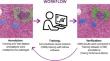
Histological assessment of autoimmune hepatitis (AIH) is challenging. As one of the possible results of these challenges, nonclassical features such as bile-duct injury stays understudied in AIH. We aim to develop a deep learning tool (artificial intelligence for autoimmune hepatitis [AI(H)]) that analyzes the liver biopsies and provides reproducible, quantifiable, and interpretable results directly from routine pathology slides. A total of 123 pre-treatment liver biopsies, whole-slide images with confirmed AIH diagnosis from the archives of the Institute of Pathology at University Hospital Basel, were used to train several convolutional neural network models in the Aiforia artificial intelligence (AI) platform. The performance of AI models was evaluated on independent test set slides against pathologist's manual annotations. The AI models were 99.4%, 88.0%, 83.9%, 81.7%, and 79.2% accurate (ratios of correct predictions) for tissue detection, liver microanatomy, necroinflammation features, bile duct damage detection, and portal inflammation detection, respectively, on hematoxylin and eosin-stained slides. Additionally, the immune cells model could detect and classify different immune cells (lymphocyte, plasma cell, macrophage, eosinophil, and neutrophil) with 72.4% accuracy. On Sirius red-stained slides, the test accuracies were 99.4%, 94.0%, and 87.6% for tissue detection, liver microanatomy, and fibrosis detection, respectively. Additionally, AI(H) showed bile duct injury in 81 AIH cases (68.6%). The AI models were found to be accurate and efficient in predicting various morphological components of AIH biopsies. The computational analysis of biopsy slides provides detailed spatial and density data of immune cells in AIH landscape, which is difficult by manual counting. AI(H) can aid in improving the reproducibility of AIH biopsy assessment and bring new descriptive and quantitative aspects to AIH histology.
期刊介绍:
Manuscripts of original studies reinforcing the evidence base of modern diagnostic pathology, using immunocytochemical, molecular and ultrastructural techniques, will be welcomed. In addition, papers on critical evaluation of diagnostic criteria but also broadsheets and guidelines with a solid evidence base will be considered. Consideration will also be given to reports of work in other fields relevant to the understanding of human pathology as well as manuscripts on the application of new methods and techniques in pathology. Submission of purely experimental articles is discouraged but manuscripts on experimental work applicable to diagnostic pathology are welcomed. Biomarker studies are welcomed but need to abide by strict rules (e.g. REMARK) of adequate sample size and relevant marker choice. Single marker studies on limited patient series without validated application will as a rule not be considered. Case reports will only be considered when they provide substantial new information with an impact on understanding disease or diagnostic practice.

 求助内容:
求助内容: 应助结果提醒方式:


