Mengjie Zeng, Flavia M Cicuttini, Anita E Wluka, Graeme Jones, Catherine L Hill, Changhai Ding, Yuanyuan Wang
{"title":"有症状的成人膝关节骨性关节炎患者内侧半月板挤压与膝关节结构恶化之间的关系--一项前瞻性队列研究。","authors":"Mengjie Zeng, Flavia M Cicuttini, Anita E Wluka, Graeme Jones, Catherine L Hill, Changhai Ding, Yuanyuan Wang","doi":"10.1007/s00256-024-04731-2","DOIUrl":null,"url":null,"abstract":"<p><strong>Objective: </strong>To examine the association between medial meniscal extrusion and structural progression in adults with symptomatic knee osteoarthritis (OA).</p><p><strong>Methods: </strong>This prospective cohort study examined 176 participants with symptomatic knee OA recruited into a randomised controlled trial. The participants underwent magnetic resonance imaging (MRI) of the study knee at baseline and approximately 2 years later. Meniscal extrusion, tibial cartilage volume, and tibiofemoral bone marrow lesions (BMLs) were measured from MRI using validated methods.</p><p><strong>Results: </strong>Participants with medial meniscal extrusion ≥ 3 mm had a higher prevalence of lateral tibiofemoral BMLs at baseline (OR = 2.21, 95% CI 1.06-4.61, p = 0.035), and those with medial meniscal extrusion 2-3 mm had a higher likelihood of lateral BML worsening over 2 years (OR = 3.76, 95% CI 1.35-10.52, p = 0.011), compared with those with medial meniscal extrusion < 2 mm. Participants with stable medial meniscal extrusion had a lower likelihood of lateral BML worsening compared with those with regression of medial meniscal extrusion over 2 years (OR = 0.20, 95% CI 0.07-0.56, p = 0.002). There were no associations between medial meniscal extrusion and tibial cartilage volume or medial tibiofemoral BMLs.</p><p><strong>Conclusions: </strong>Our study showed associations between medial meniscal extrusion and baseline prevalence and worsening over 2 years of lateral tibiofemoral BMLs in people with symptomatic knee OA. Although the reasons for the lack of associations in the medial compartment are not clear, our results suggest a role of medial meniscal extrusion in predicting structural progression in lateral knee OA and that meniscal extrusion might be a potential target in the management of knee OA.</p>","PeriodicalId":21783,"journal":{"name":"Skeletal Radiology","volume":" ","pages":"219-228"},"PeriodicalIF":1.9000,"publicationDate":"2025-02-01","publicationTypes":"Journal Article","fieldsOfStudy":null,"isOpenAccess":false,"openAccessPdf":"https://www.ncbi.nlm.nih.gov/pmc/articles/PMC11652669/pdf/","citationCount":"0","resultStr":"{\"title\":\"Association between medial meniscal extrusion and knee structural progression in adults with symptomatic knee osteoarthritis - a prospective cohort study.\",\"authors\":\"Mengjie Zeng, Flavia M Cicuttini, Anita E Wluka, Graeme Jones, Catherine L Hill, Changhai Ding, Yuanyuan Wang\",\"doi\":\"10.1007/s00256-024-04731-2\",\"DOIUrl\":null,\"url\":null,\"abstract\":\"<p><strong>Objective: </strong>To examine the association between medial meniscal extrusion and structural progression in adults with symptomatic knee osteoarthritis (OA).</p><p><strong>Methods: </strong>This prospective cohort study examined 176 participants with symptomatic knee OA recruited into a randomised controlled trial. The participants underwent magnetic resonance imaging (MRI) of the study knee at baseline and approximately 2 years later. Meniscal extrusion, tibial cartilage volume, and tibiofemoral bone marrow lesions (BMLs) were measured from MRI using validated methods.</p><p><strong>Results: </strong>Participants with medial meniscal extrusion ≥ 3 mm had a higher prevalence of lateral tibiofemoral BMLs at baseline (OR = 2.21, 95% CI 1.06-4.61, p = 0.035), and those with medial meniscal extrusion 2-3 mm had a higher likelihood of lateral BML worsening over 2 years (OR = 3.76, 95% CI 1.35-10.52, p = 0.011), compared with those with medial meniscal extrusion < 2 mm. Participants with stable medial meniscal extrusion had a lower likelihood of lateral BML worsening compared with those with regression of medial meniscal extrusion over 2 years (OR = 0.20, 95% CI 0.07-0.56, p = 0.002). There were no associations between medial meniscal extrusion and tibial cartilage volume or medial tibiofemoral BMLs.</p><p><strong>Conclusions: </strong>Our study showed associations between medial meniscal extrusion and baseline prevalence and worsening over 2 years of lateral tibiofemoral BMLs in people with symptomatic knee OA. Although the reasons for the lack of associations in the medial compartment are not clear, our results suggest a role of medial meniscal extrusion in predicting structural progression in lateral knee OA and that meniscal extrusion might be a potential target in the management of knee OA.</p>\",\"PeriodicalId\":21783,\"journal\":{\"name\":\"Skeletal Radiology\",\"volume\":\" \",\"pages\":\"219-228\"},\"PeriodicalIF\":1.9000,\"publicationDate\":\"2025-02-01\",\"publicationTypes\":\"Journal Article\",\"fieldsOfStudy\":null,\"isOpenAccess\":false,\"openAccessPdf\":\"https://www.ncbi.nlm.nih.gov/pmc/articles/PMC11652669/pdf/\",\"citationCount\":\"0\",\"resultStr\":null,\"platform\":\"Semanticscholar\",\"paperid\":null,\"PeriodicalName\":\"Skeletal Radiology\",\"FirstCategoryId\":\"3\",\"ListUrlMain\":\"https://doi.org/10.1007/s00256-024-04731-2\",\"RegionNum\":3,\"RegionCategory\":\"医学\",\"ArticlePicture\":[],\"TitleCN\":null,\"AbstractTextCN\":null,\"PMCID\":null,\"EPubDate\":\"2024/6/15 0:00:00\",\"PubModel\":\"Epub\",\"JCR\":\"Q2\",\"JCRName\":\"ORTHOPEDICS\",\"Score\":null,\"Total\":0}","platform":"Semanticscholar","paperid":null,"PeriodicalName":"Skeletal Radiology","FirstCategoryId":"3","ListUrlMain":"https://doi.org/10.1007/s00256-024-04731-2","RegionNum":3,"RegionCategory":"医学","ArticlePicture":[],"TitleCN":null,"AbstractTextCN":null,"PMCID":null,"EPubDate":"2024/6/15 0:00:00","PubModel":"Epub","JCR":"Q2","JCRName":"ORTHOPEDICS","Score":null,"Total":0}
引用次数: 0
摘要
目的研究有症状的成人膝关节骨性关节炎(OA)患者内侧半月板挤压与结构恶化之间的关系:这项前瞻性队列研究对参加随机对照试验的176名无症状膝关节OA患者进行了检查。参与者在基线和大约两年后接受了研究对象膝关节的磁共振成像(MRI)检查。磁共振成像采用经过验证的方法测量半月板挤压、胫骨软骨体积和胫股骨骨髓病变(BML):结果:与内侧半月板挤压的参与者相比,内侧半月板挤压≥3 毫米的参与者在基线时胫骨外侧骨髓病变的发生率更高(OR = 2.21,95% CI 1.06-4.61,p = 0.035);与内侧半月板挤压的参与者相比,内侧半月板挤压 2-3 毫米的参与者在 2 年内外侧骨髓病变恶化的可能性更高(OR = 3.76,95% CI 1.35-10.52,p = 0.011):我们的研究表明,在有症状的膝关节OA患者中,内侧半月板挤压与胫股外侧BML的基线发病率和2年后的恶化之间存在关联。虽然内侧区缺乏相关性的原因尚不清楚,但我们的研究结果表明,内侧半月板挤压在预测外侧膝关节OA的结构性进展中起着一定的作用,半月板挤压可能是膝关节OA治疗的一个潜在目标。
Association between medial meniscal extrusion and knee structural progression in adults with symptomatic knee osteoarthritis - a prospective cohort study.
Objective: To examine the association between medial meniscal extrusion and structural progression in adults with symptomatic knee osteoarthritis (OA).
Methods: This prospective cohort study examined 176 participants with symptomatic knee OA recruited into a randomised controlled trial. The participants underwent magnetic resonance imaging (MRI) of the study knee at baseline and approximately 2 years later. Meniscal extrusion, tibial cartilage volume, and tibiofemoral bone marrow lesions (BMLs) were measured from MRI using validated methods.
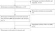
Results: Participants with medial meniscal extrusion ≥ 3 mm had a higher prevalence of lateral tibiofemoral BMLs at baseline (OR = 2.21, 95% CI 1.06-4.61, p = 0.035), and those with medial meniscal extrusion 2-3 mm had a higher likelihood of lateral BML worsening over 2 years (OR = 3.76, 95% CI 1.35-10.52, p = 0.011), compared with those with medial meniscal extrusion < 2 mm. Participants with stable medial meniscal extrusion had a lower likelihood of lateral BML worsening compared with those with regression of medial meniscal extrusion over 2 years (OR = 0.20, 95% CI 0.07-0.56, p = 0.002). There were no associations between medial meniscal extrusion and tibial cartilage volume or medial tibiofemoral BMLs.
Conclusions: Our study showed associations between medial meniscal extrusion and baseline prevalence and worsening over 2 years of lateral tibiofemoral BMLs in people with symptomatic knee OA. Although the reasons for the lack of associations in the medial compartment are not clear, our results suggest a role of medial meniscal extrusion in predicting structural progression in lateral knee OA and that meniscal extrusion might be a potential target in the management of knee OA.
期刊介绍:
Skeletal Radiology provides a forum for the dissemination of current knowledge and information dealing with disorders of the musculoskeletal system including the spine. While emphasizing the radiological aspects of the many varied skeletal abnormalities, the journal also adopts an interdisciplinary approach, reflecting the membership of the International Skeletal Society. Thus, the anatomical, pathological, physiological, clinical, metabolic and epidemiological aspects of the many entities affecting the skeleton receive appropriate consideration.
This is the Journal of the International Skeletal Society and the Official Journal of the Society of Skeletal Radiology and the Australasian Musculoskelelal Imaging Group.

 求助内容:
求助内容: 应助结果提醒方式:


