{"title":"铁蛋白沉积在 DM 引起的肝损伤中的作用","authors":"Keping Wu, Jiasi Chen, Jiawen Lin, Enyi Zhu, Xiaochang Xu, Xiuhong Yan, Lang Ju, Mingcheng Huang, Yimin Zhang","doi":"10.1007/s10534-024-00600-6","DOIUrl":null,"url":null,"abstract":"<div><p>The liver damage caused by Diabetes Mellitus (DM) has attracted increasing attention in recent years. Liver injury in DM can be caused by ferroptosis, a form of cell death caused by iron overload. However, the role of iron transporters in this context is still not clear. Herein, we attempted to shed light on the pathophysiological mechanism of ferroptosis. DM was induced in 8-week-old male rats by streptozotocin (STZ) before assessment of the degree of liver injury. Together with histopathological changes, variations in glutathione peroxidase 4 (GPX4), glutathione (GSH), superoxide dismutase (SOD), transferrin receptor 1 (TFR1), ferritin heavy chain (FTH), ferritin light chain (FTL), ferroportin and Prussian blue staining, were monitored in rat livers before and after treatment with Fer-1. In the liver of STZ-treated rats, GSH and SOD levels decreased, whereas those of malondialdehyde (MDA) increased. Expression of TFR1, FTH and FTL increased whereas that of glutathione peroxidase 4 (GPX4) and ferroportin did not change significantly. Prussian blue staining showed that iron levels increased. Histopathology showed liver fibrosis and decreased glycogen content. Fer-1 treatment reduced iron and MDA levels but GSH and SOD levels were unchanged. Expression of FTH and FTL was reduced whereas that of ferroportin showed a mild decrease. Fer-1 treatment alleviated liver fibrosis, increased glycogen content and mildly improved liver function. Our study demonstrates that ferroptosis is involved in DM-induced liver injury. Regulating the levels of iron transporters may become a new therapeutic strategy in ferroptosis-induced liver injury.</p></div>","PeriodicalId":491,"journal":{"name":"Biometals","volume":"37 5","pages":"1191 - 1200"},"PeriodicalIF":4.1000,"publicationDate":"2024-06-14","publicationTypes":"Journal Article","fieldsOfStudy":null,"isOpenAccess":false,"openAccessPdf":"","citationCount":"0","resultStr":"{\"title\":\"The role of ferroptosis in DM-induced liver injury\",\"authors\":\"Keping Wu, Jiasi Chen, Jiawen Lin, Enyi Zhu, Xiaochang Xu, Xiuhong Yan, Lang Ju, Mingcheng Huang, Yimin Zhang\",\"doi\":\"10.1007/s10534-024-00600-6\",\"DOIUrl\":null,\"url\":null,\"abstract\":\"<div><p>The liver damage caused by Diabetes Mellitus (DM) has attracted increasing attention in recent years. Liver injury in DM can be caused by ferroptosis, a form of cell death caused by iron overload. However, the role of iron transporters in this context is still not clear. Herein, we attempted to shed light on the pathophysiological mechanism of ferroptosis. DM was induced in 8-week-old male rats by streptozotocin (STZ) before assessment of the degree of liver injury. Together with histopathological changes, variations in glutathione peroxidase 4 (GPX4), glutathione (GSH), superoxide dismutase (SOD), transferrin receptor 1 (TFR1), ferritin heavy chain (FTH), ferritin light chain (FTL), ferroportin and Prussian blue staining, were monitored in rat livers before and after treatment with Fer-1. In the liver of STZ-treated rats, GSH and SOD levels decreased, whereas those of malondialdehyde (MDA) increased. Expression of TFR1, FTH and FTL increased whereas that of glutathione peroxidase 4 (GPX4) and ferroportin did not change significantly. Prussian blue staining showed that iron levels increased. Histopathology showed liver fibrosis and decreased glycogen content. Fer-1 treatment reduced iron and MDA levels but GSH and SOD levels were unchanged. Expression of FTH and FTL was reduced whereas that of ferroportin showed a mild decrease. Fer-1 treatment alleviated liver fibrosis, increased glycogen content and mildly improved liver function. Our study demonstrates that ferroptosis is involved in DM-induced liver injury. Regulating the levels of iron transporters may become a new therapeutic strategy in ferroptosis-induced liver injury.</p></div>\",\"PeriodicalId\":491,\"journal\":{\"name\":\"Biometals\",\"volume\":\"37 5\",\"pages\":\"1191 - 1200\"},\"PeriodicalIF\":4.1000,\"publicationDate\":\"2024-06-14\",\"publicationTypes\":\"Journal Article\",\"fieldsOfStudy\":null,\"isOpenAccess\":false,\"openAccessPdf\":\"\",\"citationCount\":\"0\",\"resultStr\":null,\"platform\":\"Semanticscholar\",\"paperid\":null,\"PeriodicalName\":\"Biometals\",\"FirstCategoryId\":\"99\",\"ListUrlMain\":\"https://link.springer.com/article/10.1007/s10534-024-00600-6\",\"RegionNum\":3,\"RegionCategory\":\"生物学\",\"ArticlePicture\":[],\"TitleCN\":null,\"AbstractTextCN\":null,\"PMCID\":null,\"EPubDate\":\"\",\"PubModel\":\"\",\"JCR\":\"Q2\",\"JCRName\":\"BIOCHEMISTRY & MOLECULAR BIOLOGY\",\"Score\":null,\"Total\":0}","platform":"Semanticscholar","paperid":null,"PeriodicalName":"Biometals","FirstCategoryId":"99","ListUrlMain":"https://link.springer.com/article/10.1007/s10534-024-00600-6","RegionNum":3,"RegionCategory":"生物学","ArticlePicture":[],"TitleCN":null,"AbstractTextCN":null,"PMCID":null,"EPubDate":"","PubModel":"","JCR":"Q2","JCRName":"BIOCHEMISTRY & MOLECULAR BIOLOGY","Score":null,"Total":0}
The role of ferroptosis in DM-induced liver injury
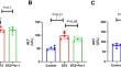
The liver damage caused by Diabetes Mellitus (DM) has attracted increasing attention in recent years. Liver injury in DM can be caused by ferroptosis, a form of cell death caused by iron overload. However, the role of iron transporters in this context is still not clear. Herein, we attempted to shed light on the pathophysiological mechanism of ferroptosis. DM was induced in 8-week-old male rats by streptozotocin (STZ) before assessment of the degree of liver injury. Together with histopathological changes, variations in glutathione peroxidase 4 (GPX4), glutathione (GSH), superoxide dismutase (SOD), transferrin receptor 1 (TFR1), ferritin heavy chain (FTH), ferritin light chain (FTL), ferroportin and Prussian blue staining, were monitored in rat livers before and after treatment with Fer-1. In the liver of STZ-treated rats, GSH and SOD levels decreased, whereas those of malondialdehyde (MDA) increased. Expression of TFR1, FTH and FTL increased whereas that of glutathione peroxidase 4 (GPX4) and ferroportin did not change significantly. Prussian blue staining showed that iron levels increased. Histopathology showed liver fibrosis and decreased glycogen content. Fer-1 treatment reduced iron and MDA levels but GSH and SOD levels were unchanged. Expression of FTH and FTL was reduced whereas that of ferroportin showed a mild decrease. Fer-1 treatment alleviated liver fibrosis, increased glycogen content and mildly improved liver function. Our study demonstrates that ferroptosis is involved in DM-induced liver injury. Regulating the levels of iron transporters may become a new therapeutic strategy in ferroptosis-induced liver injury.
期刊介绍:
BioMetals is the only established journal to feature the important role of metal ions in chemistry, biology, biochemistry, environmental science, and medicine. BioMetals is an international, multidisciplinary journal singularly devoted to the rapid publication of the fundamental advances of both basic and applied research in this field. BioMetals offers a forum for innovative research and clinical results on the structure and function of:
- metal ions
- metal chelates,
- siderophores,
- metal-containing proteins
- biominerals in all biosystems.
- BioMetals rapidly publishes original articles and reviews.
BioMetals is a journal for metals researchers who practice in medicine, biochemistry, pharmacology, toxicology, microbiology, cell biology, chemistry, and plant physiology who are based academic, industrial and government laboratories.
 求助内容:
求助内容: 应助结果提醒方式:


