André S B Oliveira, Luciano C P C Leonel, Megan M J Bauman, Alessandro De Bonis, Edward R LaHood, Stephen Graepel, Michael J Link, Carlos D Pinheiro-Neto, Nirusha Lachman, Jonathan M Morris, Maria Peris-Celda
{"title":"用于神经解剖学教育的摄影测量扫描--新型多摄像头系统:技术说明。","authors":"André S B Oliveira, Luciano C P C Leonel, Megan M J Bauman, Alessandro De Bonis, Edward R LaHood, Stephen Graepel, Michael J Link, Carlos D Pinheiro-Neto, Nirusha Lachman, Jonathan M Morris, Maria Peris-Celda","doi":"10.1007/s12021-024-09672-8","DOIUrl":null,"url":null,"abstract":"<p><p>Photogrammetry scans has directed attention to the development of advanced camera systems to improve the creation of three-dimensional (3D) models, especially for educational and medical-related purposes. This could be a potential cost-effective method for neuroanatomy education, especially when access to laboratory-based learning is limited. The aim of this study was to describe a new photogrammetry system based on a 5 Digital Single-Lens Reflex (DSLR) cameras setup to optimize accuracy of neuroanatomical 3D models. One formalin-fixed brain and specimen and one dry skull were used for dissections and scanning using the photogrammetry technique. After each dissection, the specimens were placed inside a new MedCreator® scanner (MedReality, Thyng, Chicago, IL) to be scanned with the final 3D model being displayed on SketchFab® (Epic, Cary, NC) and MedReality® platforms. The scanner consisted of 5 cameras arranged vertically facing the specimen, which was positioned on a platform in the center of the scanner. The new multi-camera system contains automated software packages, which allowed for quick rendering and creation of a high-quality 3D models. Following uploading the 3D models to the SketchFab® and MedReality® platforms for display, the models can be freely manipulated in various angles and magnifications in any devices free of charge for users. Therefore, photogrammetry scans with this new multi-camera system have the potential to enhance the accuracy and resolution of the 3D models, along with shortening creation time of the models. This system can serve as an important tool to optimize neuroanatomy education and ultimately, improve patient outcomes.</p>","PeriodicalId":49761,"journal":{"name":"Neuroinformatics","volume":" ","pages":"317-327"},"PeriodicalIF":3.1000,"publicationDate":"2024-07-01","publicationTypes":"Journal Article","fieldsOfStudy":null,"isOpenAccess":false,"openAccessPdf":"","citationCount":"0","resultStr":"{\"title\":\"Photogrammetry scans for neuroanatomy education - a new multi-camera system: technical note.\",\"authors\":\"André S B Oliveira, Luciano C P C Leonel, Megan M J Bauman, Alessandro De Bonis, Edward R LaHood, Stephen Graepel, Michael J Link, Carlos D Pinheiro-Neto, Nirusha Lachman, Jonathan M Morris, Maria Peris-Celda\",\"doi\":\"10.1007/s12021-024-09672-8\",\"DOIUrl\":null,\"url\":null,\"abstract\":\"<p><p>Photogrammetry scans has directed attention to the development of advanced camera systems to improve the creation of three-dimensional (3D) models, especially for educational and medical-related purposes. This could be a potential cost-effective method for neuroanatomy education, especially when access to laboratory-based learning is limited. The aim of this study was to describe a new photogrammetry system based on a 5 Digital Single-Lens Reflex (DSLR) cameras setup to optimize accuracy of neuroanatomical 3D models. One formalin-fixed brain and specimen and one dry skull were used for dissections and scanning using the photogrammetry technique. After each dissection, the specimens were placed inside a new MedCreator® scanner (MedReality, Thyng, Chicago, IL) to be scanned with the final 3D model being displayed on SketchFab® (Epic, Cary, NC) and MedReality® platforms. The scanner consisted of 5 cameras arranged vertically facing the specimen, which was positioned on a platform in the center of the scanner. The new multi-camera system contains automated software packages, which allowed for quick rendering and creation of a high-quality 3D models. Following uploading the 3D models to the SketchFab® and MedReality® platforms for display, the models can be freely manipulated in various angles and magnifications in any devices free of charge for users. Therefore, photogrammetry scans with this new multi-camera system have the potential to enhance the accuracy and resolution of the 3D models, along with shortening creation time of the models. This system can serve as an important tool to optimize neuroanatomy education and ultimately, improve patient outcomes.</p>\",\"PeriodicalId\":49761,\"journal\":{\"name\":\"Neuroinformatics\",\"volume\":\" \",\"pages\":\"317-327\"},\"PeriodicalIF\":3.1000,\"publicationDate\":\"2024-07-01\",\"publicationTypes\":\"Journal Article\",\"fieldsOfStudy\":null,\"isOpenAccess\":false,\"openAccessPdf\":\"\",\"citationCount\":\"0\",\"resultStr\":null,\"platform\":\"Semanticscholar\",\"paperid\":null,\"PeriodicalName\":\"Neuroinformatics\",\"FirstCategoryId\":\"3\",\"ListUrlMain\":\"https://doi.org/10.1007/s12021-024-09672-8\",\"RegionNum\":4,\"RegionCategory\":\"医学\",\"ArticlePicture\":[],\"TitleCN\":null,\"AbstractTextCN\":null,\"PMCID\":null,\"EPubDate\":\"2024/6/13 0:00:00\",\"PubModel\":\"Epub\",\"JCR\":\"Q2\",\"JCRName\":\"COMPUTER SCIENCE, INTERDISCIPLINARY APPLICATIONS\",\"Score\":null,\"Total\":0}","platform":"Semanticscholar","paperid":null,"PeriodicalName":"Neuroinformatics","FirstCategoryId":"3","ListUrlMain":"https://doi.org/10.1007/s12021-024-09672-8","RegionNum":4,"RegionCategory":"医学","ArticlePicture":[],"TitleCN":null,"AbstractTextCN":null,"PMCID":null,"EPubDate":"2024/6/13 0:00:00","PubModel":"Epub","JCR":"Q2","JCRName":"COMPUTER SCIENCE, INTERDISCIPLINARY APPLICATIONS","Score":null,"Total":0}
Photogrammetry scans for neuroanatomy education - a new multi-camera system: technical note.
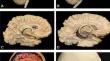
Photogrammetry scans has directed attention to the development of advanced camera systems to improve the creation of three-dimensional (3D) models, especially for educational and medical-related purposes. This could be a potential cost-effective method for neuroanatomy education, especially when access to laboratory-based learning is limited. The aim of this study was to describe a new photogrammetry system based on a 5 Digital Single-Lens Reflex (DSLR) cameras setup to optimize accuracy of neuroanatomical 3D models. One formalin-fixed brain and specimen and one dry skull were used for dissections and scanning using the photogrammetry technique. After each dissection, the specimens were placed inside a new MedCreator® scanner (MedReality, Thyng, Chicago, IL) to be scanned with the final 3D model being displayed on SketchFab® (Epic, Cary, NC) and MedReality® platforms. The scanner consisted of 5 cameras arranged vertically facing the specimen, which was positioned on a platform in the center of the scanner. The new multi-camera system contains automated software packages, which allowed for quick rendering and creation of a high-quality 3D models. Following uploading the 3D models to the SketchFab® and MedReality® platforms for display, the models can be freely manipulated in various angles and magnifications in any devices free of charge for users. Therefore, photogrammetry scans with this new multi-camera system have the potential to enhance the accuracy and resolution of the 3D models, along with shortening creation time of the models. This system can serve as an important tool to optimize neuroanatomy education and ultimately, improve patient outcomes.
期刊介绍:
Neuroinformatics publishes original articles and reviews with an emphasis on data structure and software tools related to analysis, modeling, integration, and sharing in all areas of neuroscience research. The editors particularly invite contributions on: (1) Theory and methodology, including discussions on ontologies, modeling approaches, database design, and meta-analyses; (2) Descriptions of developed databases and software tools, and of the methods for their distribution; (3) Relevant experimental results, such as reports accompanie by the release of massive data sets; (4) Computational simulations of models integrating and organizing complex data; and (5) Neuroengineering approaches, including hardware, robotics, and information theory studies.
 求助内容:
求助内容: 应助结果提醒方式:


