Shan Lv, Hongfei Tai, Jun Sun, Zhizheng Zhuo, Yunyun Duan, Shaocheng Liu, An Wang, Zaiqiang Zhang, Yaou Liu
{"title":"绘制神经元核内包涵体病患者大脑宏观和微观结构改变的图谱。","authors":"Shan Lv, Hongfei Tai, Jun Sun, Zhizheng Zhuo, Yunyun Duan, Shaocheng Liu, An Wang, Zaiqiang Zhang, Yaou Liu","doi":"10.1007/s00234-024-03406-y","DOIUrl":null,"url":null,"abstract":"<p><strong>Background and purpose: </strong>Neuronal intranuclear inclusion disease (NIID) is a rare complex neurodegenerative disorder presents with various radiological features. The study aimed to investigate the structural abnormalities in NIID using multi-shell diffusion MR.</p><p><strong>Materials and methods: </strong>Twenty-eight patients with adult-onset NIID and 32 healthy controls were included. Volumetric and diffusion MRI measures, including volume, fractional anisotropy (FA), mean diffusivity (MD), intracellular volume fraction (ICVF), orientation dispersion index (ODI), and isotropic volume fraction (ISOVF) of six brain structures, including cortex, subcortical GM, cerebral WM, cerebellar GM and WM, and brainstem, were obtained and compared between NIID and healthy controls. Associations between MRI measures and clinical variables were investigated.</p><p><strong>Results: </strong>Brain lesions of NIID included corticomedullary junction lesions on DWI, confluent leukoencephalopathy, lesions on callosum, cerebellar middle peduncle, cerebellar paravermal area and brainstem, and brain atrophy. Compared to healthy controls, NIID showed extensive volume loss of all the six brain regions (all p < 0.001); lower FA in cerebral WM (p < 0.001); higher MD in all WM regions; lower ODI in cortex (p < 0.001); higher ODI in subcortical GM (p < 0.001) and brainstem (p = 0.016); lower ICVF in brainstem (p = 0.001), and cerebral WM (p < 0.001); higher ISOVF in all the brain regions (p < 0.001). Higher MD of cerebellar WM was associated with worse cognitive level as evaluated by MoCA scores (p = 0.011).</p><p><strong>Conclusions: </strong>NIID patients demonstrated widespread brain atrophy but heterogeneous diffusion alterations. Cerebellar WM integrity impairment was correlated with the cognitive decline. The findings of the current study offer a sophisticated picture of brain structural alterations in NIID.</p>","PeriodicalId":19422,"journal":{"name":"Neuroradiology","volume":" ","pages":"1373-1382"},"PeriodicalIF":2.4000,"publicationDate":"2024-08-01","publicationTypes":"Journal Article","fieldsOfStudy":null,"isOpenAccess":false,"openAccessPdf":"","citationCount":"0","resultStr":"{\"title\":\"Mapping macrostructural and microstructural brain alterations in patients with neuronal intranuclear inclusion disease.\",\"authors\":\"Shan Lv, Hongfei Tai, Jun Sun, Zhizheng Zhuo, Yunyun Duan, Shaocheng Liu, An Wang, Zaiqiang Zhang, Yaou Liu\",\"doi\":\"10.1007/s00234-024-03406-y\",\"DOIUrl\":null,\"url\":null,\"abstract\":\"<p><strong>Background and purpose: </strong>Neuronal intranuclear inclusion disease (NIID) is a rare complex neurodegenerative disorder presents with various radiological features. The study aimed to investigate the structural abnormalities in NIID using multi-shell diffusion MR.</p><p><strong>Materials and methods: </strong>Twenty-eight patients with adult-onset NIID and 32 healthy controls were included. Volumetric and diffusion MRI measures, including volume, fractional anisotropy (FA), mean diffusivity (MD), intracellular volume fraction (ICVF), orientation dispersion index (ODI), and isotropic volume fraction (ISOVF) of six brain structures, including cortex, subcortical GM, cerebral WM, cerebellar GM and WM, and brainstem, were obtained and compared between NIID and healthy controls. Associations between MRI measures and clinical variables were investigated.</p><p><strong>Results: </strong>Brain lesions of NIID included corticomedullary junction lesions on DWI, confluent leukoencephalopathy, lesions on callosum, cerebellar middle peduncle, cerebellar paravermal area and brainstem, and brain atrophy. Compared to healthy controls, NIID showed extensive volume loss of all the six brain regions (all p < 0.001); lower FA in cerebral WM (p < 0.001); higher MD in all WM regions; lower ODI in cortex (p < 0.001); higher ODI in subcortical GM (p < 0.001) and brainstem (p = 0.016); lower ICVF in brainstem (p = 0.001), and cerebral WM (p < 0.001); higher ISOVF in all the brain regions (p < 0.001). Higher MD of cerebellar WM was associated with worse cognitive level as evaluated by MoCA scores (p = 0.011).</p><p><strong>Conclusions: </strong>NIID patients demonstrated widespread brain atrophy but heterogeneous diffusion alterations. Cerebellar WM integrity impairment was correlated with the cognitive decline. The findings of the current study offer a sophisticated picture of brain structural alterations in NIID.</p>\",\"PeriodicalId\":19422,\"journal\":{\"name\":\"Neuroradiology\",\"volume\":\" \",\"pages\":\"1373-1382\"},\"PeriodicalIF\":2.4000,\"publicationDate\":\"2024-08-01\",\"publicationTypes\":\"Journal Article\",\"fieldsOfStudy\":null,\"isOpenAccess\":false,\"openAccessPdf\":\"\",\"citationCount\":\"0\",\"resultStr\":null,\"platform\":\"Semanticscholar\",\"paperid\":null,\"PeriodicalName\":\"Neuroradiology\",\"FirstCategoryId\":\"3\",\"ListUrlMain\":\"https://doi.org/10.1007/s00234-024-03406-y\",\"RegionNum\":3,\"RegionCategory\":\"医学\",\"ArticlePicture\":[],\"TitleCN\":null,\"AbstractTextCN\":null,\"PMCID\":null,\"EPubDate\":\"2024/6/13 0:00:00\",\"PubModel\":\"Epub\",\"JCR\":\"Q2\",\"JCRName\":\"CLINICAL NEUROLOGY\",\"Score\":null,\"Total\":0}","platform":"Semanticscholar","paperid":null,"PeriodicalName":"Neuroradiology","FirstCategoryId":"3","ListUrlMain":"https://doi.org/10.1007/s00234-024-03406-y","RegionNum":3,"RegionCategory":"医学","ArticlePicture":[],"TitleCN":null,"AbstractTextCN":null,"PMCID":null,"EPubDate":"2024/6/13 0:00:00","PubModel":"Epub","JCR":"Q2","JCRName":"CLINICAL NEUROLOGY","Score":null,"Total":0}
Mapping macrostructural and microstructural brain alterations in patients with neuronal intranuclear inclusion disease.
Background and purpose: Neuronal intranuclear inclusion disease (NIID) is a rare complex neurodegenerative disorder presents with various radiological features. The study aimed to investigate the structural abnormalities in NIID using multi-shell diffusion MR.
Materials and methods: Twenty-eight patients with adult-onset NIID and 32 healthy controls were included. Volumetric and diffusion MRI measures, including volume, fractional anisotropy (FA), mean diffusivity (MD), intracellular volume fraction (ICVF), orientation dispersion index (ODI), and isotropic volume fraction (ISOVF) of six brain structures, including cortex, subcortical GM, cerebral WM, cerebellar GM and WM, and brainstem, were obtained and compared between NIID and healthy controls. Associations between MRI measures and clinical variables were investigated.
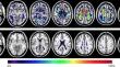
Results: Brain lesions of NIID included corticomedullary junction lesions on DWI, confluent leukoencephalopathy, lesions on callosum, cerebellar middle peduncle, cerebellar paravermal area and brainstem, and brain atrophy. Compared to healthy controls, NIID showed extensive volume loss of all the six brain regions (all p < 0.001); lower FA in cerebral WM (p < 0.001); higher MD in all WM regions; lower ODI in cortex (p < 0.001); higher ODI in subcortical GM (p < 0.001) and brainstem (p = 0.016); lower ICVF in brainstem (p = 0.001), and cerebral WM (p < 0.001); higher ISOVF in all the brain regions (p < 0.001). Higher MD of cerebellar WM was associated with worse cognitive level as evaluated by MoCA scores (p = 0.011).
Conclusions: NIID patients demonstrated widespread brain atrophy but heterogeneous diffusion alterations. Cerebellar WM integrity impairment was correlated with the cognitive decline. The findings of the current study offer a sophisticated picture of brain structural alterations in NIID.
期刊介绍:
Neuroradiology aims to provide state-of-the-art medical and scientific information in the fields of Neuroradiology, Neurosciences, Neurology, Psychiatry, Neurosurgery, and related medical specialities. Neuroradiology as the official Journal of the European Society of Neuroradiology receives submissions from all parts of the world and publishes peer-reviewed original research, comprehensive reviews, educational papers, opinion papers, and short reports on exceptional clinical observations and new technical developments in the field of Neuroimaging and Neurointervention. The journal has subsections for Diagnostic and Interventional Neuroradiology, Advanced Neuroimaging, Paediatric Neuroradiology, Head-Neck-ENT Radiology, Spine Neuroradiology, and for submissions from Japan. Neuroradiology aims to provide new knowledge about and insights into the function and pathology of the human nervous system that may help to better diagnose and treat nervous system diseases. Neuroradiology is a member of the Committee on Publication Ethics (COPE) and follows the COPE core practices. Neuroradiology prefers articles that are free of bias, self-critical regarding limitations, transparent and clear in describing study participants, methods, and statistics, and short in presenting results. Before peer-review all submissions are automatically checked by iThenticate to assess for potential overlap in prior publication.

 求助内容:
求助内容: 应助结果提醒方式:


