{"title":"预测自发性脑内出血血肿扩大的机器学习:系统综述和荟萃分析。","authors":"Yihua Liu, Fengfeng Zhao, Enjing Niu, Liang Chen","doi":"10.1007/s00234-024-03399-8","DOIUrl":null,"url":null,"abstract":"<p><strong>Purpose: </strong>Early identification of hematoma enlargement and persistent hematoma expansion (HE) in patients with cerebral hemorrhage is increasingly crucial for determining clinical treatments. However, due to the lack of clinically effective tools, radiomics has been gradually introduced into the early identification of hematoma enlargement. Though, radiomics has limited predictive accuracy due to variations in procedures. Therefore, we conducted a systematic review and meta-analysis to explore the value of radiomics in the early detection of HE in patients with cerebral hemorrhage.</p><p><strong>Methods: </strong>Eligible studies were systematically searched in PubMed, Embase, Cochrane and Web of Science from inception to April 8, 2024. English articles are considered eligible. The radiomics quality scoring (RQS) tool was used to evaluate included studies.</p><p><strong>Results: </strong>A total of 34 studies were identified with sample sizes ranging from 108 to 3016. Eleven types of models were involved, and the types of modeling contained mainly clinical, radiomic, and radiomic plus clinical features. The radiomics models seem to have better performance (0.77 and 0.73 C-index in the training cohort and validation cohort, respectively) than the clinical models (0.69 C-index in the training cohort and 0.70 C-index in the validation cohort) in discriminating HE. However, the C-index was the highest for the combined model in both the training (0.82) and validation (0.79) cohorts.</p><p><strong>Conclusions: </strong>Machine learning based on radiomic plus clinical features has the best predictive performance for HE, followed by machine learning based on radiomic features, and can be used as a potential tool to assist clinicians in early judgment.</p>","PeriodicalId":19422,"journal":{"name":"Neuroradiology","volume":null,"pages":null},"PeriodicalIF":2.4000,"publicationDate":"2024-09-01","publicationTypes":"Journal Article","fieldsOfStudy":null,"isOpenAccess":false,"openAccessPdf":"","citationCount":"0","resultStr":"{\"title\":\"Machine learning for predicting hematoma expansion in spontaneous intracerebral hemorrhage: a systematic review and meta-analysis.\",\"authors\":\"Yihua Liu, Fengfeng Zhao, Enjing Niu, Liang Chen\",\"doi\":\"10.1007/s00234-024-03399-8\",\"DOIUrl\":null,\"url\":null,\"abstract\":\"<p><strong>Purpose: </strong>Early identification of hematoma enlargement and persistent hematoma expansion (HE) in patients with cerebral hemorrhage is increasingly crucial for determining clinical treatments. However, due to the lack of clinically effective tools, radiomics has been gradually introduced into the early identification of hematoma enlargement. Though, radiomics has limited predictive accuracy due to variations in procedures. Therefore, we conducted a systematic review and meta-analysis to explore the value of radiomics in the early detection of HE in patients with cerebral hemorrhage.</p><p><strong>Methods: </strong>Eligible studies were systematically searched in PubMed, Embase, Cochrane and Web of Science from inception to April 8, 2024. English articles are considered eligible. The radiomics quality scoring (RQS) tool was used to evaluate included studies.</p><p><strong>Results: </strong>A total of 34 studies were identified with sample sizes ranging from 108 to 3016. Eleven types of models were involved, and the types of modeling contained mainly clinical, radiomic, and radiomic plus clinical features. The radiomics models seem to have better performance (0.77 and 0.73 C-index in the training cohort and validation cohort, respectively) than the clinical models (0.69 C-index in the training cohort and 0.70 C-index in the validation cohort) in discriminating HE. However, the C-index was the highest for the combined model in both the training (0.82) and validation (0.79) cohorts.</p><p><strong>Conclusions: </strong>Machine learning based on radiomic plus clinical features has the best predictive performance for HE, followed by machine learning based on radiomic features, and can be used as a potential tool to assist clinicians in early judgment.</p>\",\"PeriodicalId\":19422,\"journal\":{\"name\":\"Neuroradiology\",\"volume\":null,\"pages\":null},\"PeriodicalIF\":2.4000,\"publicationDate\":\"2024-09-01\",\"publicationTypes\":\"Journal Article\",\"fieldsOfStudy\":null,\"isOpenAccess\":false,\"openAccessPdf\":\"\",\"citationCount\":\"0\",\"resultStr\":null,\"platform\":\"Semanticscholar\",\"paperid\":null,\"PeriodicalName\":\"Neuroradiology\",\"FirstCategoryId\":\"3\",\"ListUrlMain\":\"https://doi.org/10.1007/s00234-024-03399-8\",\"RegionNum\":3,\"RegionCategory\":\"医学\",\"ArticlePicture\":[],\"TitleCN\":null,\"AbstractTextCN\":null,\"PMCID\":null,\"EPubDate\":\"2024/6/12 0:00:00\",\"PubModel\":\"Epub\",\"JCR\":\"Q2\",\"JCRName\":\"CLINICAL NEUROLOGY\",\"Score\":null,\"Total\":0}","platform":"Semanticscholar","paperid":null,"PeriodicalName":"Neuroradiology","FirstCategoryId":"3","ListUrlMain":"https://doi.org/10.1007/s00234-024-03399-8","RegionNum":3,"RegionCategory":"医学","ArticlePicture":[],"TitleCN":null,"AbstractTextCN":null,"PMCID":null,"EPubDate":"2024/6/12 0:00:00","PubModel":"Epub","JCR":"Q2","JCRName":"CLINICAL NEUROLOGY","Score":null,"Total":0}
引用次数: 0
摘要
目的:脑出血患者血肿扩大和持续性血肿扩大(HE)的早期识别对于决定临床治疗越来越重要。然而,由于缺乏临床有效的工具,放射组学已逐渐被引入血肿扩大的早期识别中。尽管如此,由于操作程序的不同,放射组学的预测准确性有限。因此,我们进行了一项系统性回顾和荟萃分析,以探讨放射组学在早期发现脑出血患者 HE 中的价值:方法:在 PubMed、Embase、Cochrane 和 Web of Science 中系统检索了从开始到 2024 年 4 月 8 日的符合条件的研究。符合条件的文章均为英文文章。采用放射组学质量评分(RQS)工具对纳入的研究进行评估:共确定了 34 项研究,样本量从 108 到 3016 不等。共涉及 11 种模型,模型类型主要包括临床特征、放射组学特征和放射组学加临床特征。放射组学模型在鉴别 HE 方面的表现(训练队列和验证队列中的 C 指数分别为 0.77 和 0.73)似乎优于临床模型(训练队列中的 C 指数为 0.69,验证队列中的 C 指数为 0.70)。然而,在训练队列(0.82)和验证队列(0.79)中,组合模型的 C 指数都是最高的:结论:基于放射学和临床特征的机器学习对 HE 的预测效果最好,其次是基于放射学特征的机器学习,可作为辅助临床医生进行早期判断的潜在工具。
Machine learning for predicting hematoma expansion in spontaneous intracerebral hemorrhage: a systematic review and meta-analysis.
Purpose: Early identification of hematoma enlargement and persistent hematoma expansion (HE) in patients with cerebral hemorrhage is increasingly crucial for determining clinical treatments. However, due to the lack of clinically effective tools, radiomics has been gradually introduced into the early identification of hematoma enlargement. Though, radiomics has limited predictive accuracy due to variations in procedures. Therefore, we conducted a systematic review and meta-analysis to explore the value of radiomics in the early detection of HE in patients with cerebral hemorrhage.
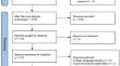
Methods: Eligible studies were systematically searched in PubMed, Embase, Cochrane and Web of Science from inception to April 8, 2024. English articles are considered eligible. The radiomics quality scoring (RQS) tool was used to evaluate included studies.
Results: A total of 34 studies were identified with sample sizes ranging from 108 to 3016. Eleven types of models were involved, and the types of modeling contained mainly clinical, radiomic, and radiomic plus clinical features. The radiomics models seem to have better performance (0.77 and 0.73 C-index in the training cohort and validation cohort, respectively) than the clinical models (0.69 C-index in the training cohort and 0.70 C-index in the validation cohort) in discriminating HE. However, the C-index was the highest for the combined model in both the training (0.82) and validation (0.79) cohorts.
Conclusions: Machine learning based on radiomic plus clinical features has the best predictive performance for HE, followed by machine learning based on radiomic features, and can be used as a potential tool to assist clinicians in early judgment.
期刊介绍:
Neuroradiology aims to provide state-of-the-art medical and scientific information in the fields of Neuroradiology, Neurosciences, Neurology, Psychiatry, Neurosurgery, and related medical specialities. Neuroradiology as the official Journal of the European Society of Neuroradiology receives submissions from all parts of the world and publishes peer-reviewed original research, comprehensive reviews, educational papers, opinion papers, and short reports on exceptional clinical observations and new technical developments in the field of Neuroimaging and Neurointervention. The journal has subsections for Diagnostic and Interventional Neuroradiology, Advanced Neuroimaging, Paediatric Neuroradiology, Head-Neck-ENT Radiology, Spine Neuroradiology, and for submissions from Japan. Neuroradiology aims to provide new knowledge about and insights into the function and pathology of the human nervous system that may help to better diagnose and treat nervous system diseases. Neuroradiology is a member of the Committee on Publication Ethics (COPE) and follows the COPE core practices. Neuroradiology prefers articles that are free of bias, self-critical regarding limitations, transparent and clear in describing study participants, methods, and statistics, and short in presenting results. Before peer-review all submissions are automatically checked by iThenticate to assess for potential overlap in prior publication.

 求助内容:
求助内容: 应助结果提醒方式:


