Mihaela Daniela Manta, Mugurel Constantin Rusu, Sorin Hostiuc, Răzvan Costin Tudose, Bogdan Adrian Manta, Adelina Maria Jianu
{"title":"颈动脉分叉的垂直地形图--原始研究和综述。","authors":"Mihaela Daniela Manta, Mugurel Constantin Rusu, Sorin Hostiuc, Răzvan Costin Tudose, Bogdan Adrian Manta, Adelina Maria Jianu","doi":"10.1007/s00276-024-03404-y","DOIUrl":null,"url":null,"abstract":"<p><strong>Purpose: </strong>The vertical level of carotid bifurcation (CB) is commonly indicated at the superior margin of the thyroid cartilage. Few studies observed the CB vertical topography. It was aimed at studying the vertical location of the CB as referred to vertebral and anterior cervical landmarks.</p><p><strong>Methods: </strong>An archived lot of 147 computed tomography angiograms was documented for the vertical level of CB referred to vertebral and anterior cervical landmarks. The topography of the CB in relation to anterior landmarks was classified into seven types: (1) at the superior margin of the thyroid cartilage; (2) between the hyoid and the thyroid cartilage; (3) at the hyoid level; (4) between the hyoid and mandible; (5) subgonial or supragonial CB; (6) lower cervical level; (7) intrathoracic.</p><p><strong>Results: </strong>The most common locations of CB were at C3 (27.21%), C3/C4 (26.19%) and C4 (25.51%). Bilateral symmetry of CB was found in 51.7%, except for C2 and C5/C6. Type 7 was not found, type 3 occurred in 39.12%, type 2 in 24.49%, type 1 in 13.95%, type 4 in 13.61%, type 5 in 6.12%, and type 6 in 2.72% (294 CBs). Bilateral symmetry of anterior types was found in 59.86%. Statistically significant correlations were found between sex and both left and right types and vertebral levels of CB.</p><p><strong>Conclusions: </strong>The vertical topography of the CB is highly variable and has sex-related specificity. This detail should be included in the teaching of anatomy. Surgeons and interventionists should better document the carotid anatomy on a case-by-case basis.</p>","PeriodicalId":49461,"journal":{"name":"Surgical and Radiologic Anatomy","volume":" ","pages":"1253-1263"},"PeriodicalIF":1.4000,"publicationDate":"2024-08-01","publicationTypes":"Journal Article","fieldsOfStudy":null,"isOpenAccess":false,"openAccessPdf":"https://www.ncbi.nlm.nih.gov/pmc/articles/PMC11246274/pdf/","citationCount":"0","resultStr":"{\"title\":\"The vertical topography of the carotid bifurcation - original study and review.\",\"authors\":\"Mihaela Daniela Manta, Mugurel Constantin Rusu, Sorin Hostiuc, Răzvan Costin Tudose, Bogdan Adrian Manta, Adelina Maria Jianu\",\"doi\":\"10.1007/s00276-024-03404-y\",\"DOIUrl\":null,\"url\":null,\"abstract\":\"<p><strong>Purpose: </strong>The vertical level of carotid bifurcation (CB) is commonly indicated at the superior margin of the thyroid cartilage. Few studies observed the CB vertical topography. It was aimed at studying the vertical location of the CB as referred to vertebral and anterior cervical landmarks.</p><p><strong>Methods: </strong>An archived lot of 147 computed tomography angiograms was documented for the vertical level of CB referred to vertebral and anterior cervical landmarks. The topography of the CB in relation to anterior landmarks was classified into seven types: (1) at the superior margin of the thyroid cartilage; (2) between the hyoid and the thyroid cartilage; (3) at the hyoid level; (4) between the hyoid and mandible; (5) subgonial or supragonial CB; (6) lower cervical level; (7) intrathoracic.</p><p><strong>Results: </strong>The most common locations of CB were at C3 (27.21%), C3/C4 (26.19%) and C4 (25.51%). Bilateral symmetry of CB was found in 51.7%, except for C2 and C5/C6. Type 7 was not found, type 3 occurred in 39.12%, type 2 in 24.49%, type 1 in 13.95%, type 4 in 13.61%, type 5 in 6.12%, and type 6 in 2.72% (294 CBs). Bilateral symmetry of anterior types was found in 59.86%. Statistically significant correlations were found between sex and both left and right types and vertebral levels of CB.</p><p><strong>Conclusions: </strong>The vertical topography of the CB is highly variable and has sex-related specificity. This detail should be included in the teaching of anatomy. Surgeons and interventionists should better document the carotid anatomy on a case-by-case basis.</p>\",\"PeriodicalId\":49461,\"journal\":{\"name\":\"Surgical and Radiologic Anatomy\",\"volume\":\" \",\"pages\":\"1253-1263\"},\"PeriodicalIF\":1.4000,\"publicationDate\":\"2024-08-01\",\"publicationTypes\":\"Journal Article\",\"fieldsOfStudy\":null,\"isOpenAccess\":false,\"openAccessPdf\":\"https://www.ncbi.nlm.nih.gov/pmc/articles/PMC11246274/pdf/\",\"citationCount\":\"0\",\"resultStr\":null,\"platform\":\"Semanticscholar\",\"paperid\":null,\"PeriodicalName\":\"Surgical and Radiologic Anatomy\",\"FirstCategoryId\":\"3\",\"ListUrlMain\":\"https://doi.org/10.1007/s00276-024-03404-y\",\"RegionNum\":4,\"RegionCategory\":\"医学\",\"ArticlePicture\":[],\"TitleCN\":null,\"AbstractTextCN\":null,\"PMCID\":null,\"EPubDate\":\"2024/6/7 0:00:00\",\"PubModel\":\"Epub\",\"JCR\":\"Q2\",\"JCRName\":\"Medicine\",\"Score\":null,\"Total\":0}","platform":"Semanticscholar","paperid":null,"PeriodicalName":"Surgical and Radiologic Anatomy","FirstCategoryId":"3","ListUrlMain":"https://doi.org/10.1007/s00276-024-03404-y","RegionNum":4,"RegionCategory":"医学","ArticlePicture":[],"TitleCN":null,"AbstractTextCN":null,"PMCID":null,"EPubDate":"2024/6/7 0:00:00","PubModel":"Epub","JCR":"Q2","JCRName":"Medicine","Score":null,"Total":0}
The vertical topography of the carotid bifurcation - original study and review.
Purpose: The vertical level of carotid bifurcation (CB) is commonly indicated at the superior margin of the thyroid cartilage. Few studies observed the CB vertical topography. It was aimed at studying the vertical location of the CB as referred to vertebral and anterior cervical landmarks.
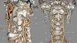
Methods: An archived lot of 147 computed tomography angiograms was documented for the vertical level of CB referred to vertebral and anterior cervical landmarks. The topography of the CB in relation to anterior landmarks was classified into seven types: (1) at the superior margin of the thyroid cartilage; (2) between the hyoid and the thyroid cartilage; (3) at the hyoid level; (4) between the hyoid and mandible; (5) subgonial or supragonial CB; (6) lower cervical level; (7) intrathoracic.
Results: The most common locations of CB were at C3 (27.21%), C3/C4 (26.19%) and C4 (25.51%). Bilateral symmetry of CB was found in 51.7%, except for C2 and C5/C6. Type 7 was not found, type 3 occurred in 39.12%, type 2 in 24.49%, type 1 in 13.95%, type 4 in 13.61%, type 5 in 6.12%, and type 6 in 2.72% (294 CBs). Bilateral symmetry of anterior types was found in 59.86%. Statistically significant correlations were found between sex and both left and right types and vertebral levels of CB.
Conclusions: The vertical topography of the CB is highly variable and has sex-related specificity. This detail should be included in the teaching of anatomy. Surgeons and interventionists should better document the carotid anatomy on a case-by-case basis.
期刊介绍:
Anatomy is a morphological science which cannot fail to interest the clinician. The practical application of anatomical research to clinical problems necessitates special adaptation and selectivity in choosing from numerous international works. Although there is a tendency to believe that meaningful advances in anatomy are unlikely, constant revision is necessary. Surgical and Radiologic Anatomy, the first international journal of Clinical anatomy has been created in this spirit.
Its goal is to serve clinicians, regardless of speciality-physicians, surgeons, radiologists or other specialists-as an indispensable aid with which they can improve their knowledge of anatomy. Each issue includes: Original papers, review articles, articles on the anatomical bases of medical, surgical and radiological techniques, articles of normal radiologic anatomy, brief reviews of anatomical publications of clinical interest.
Particular attention is given to high quality illustrations, which are indispensable for a better understanding of anatomical problems.
Surgical and Radiologic Anatomy is a journal written by anatomists for clinicians with a special interest in anatomy.

 求助内容:
求助内容: 应助结果提醒方式:


