{"title":"研究用二氧化钛掺杂氧化石墨烯纳米粒子合成的真菌提取物的生物活性。","authors":"K. Kaviyarasu","doi":"10.1002/jemt.24625","DOIUrl":null,"url":null,"abstract":"<div>\n \n \n <section>\n \n <p>Nanoparticles of titanium dioxide (TiO<sub>2</sub>) were made by reacting graphene oxide (GO) with <i>Lawsonia inermis</i> leaf extract. X-ray diffraction (XRD) analysis revealed crystalline TiO<sub>2</sub> doped GO nanoparticles composed of a variety of anatase phases. Initially, UV–vis spectroscopy was performed to confirm the biogenesis of TiO<sub>2</sub> doped GO nanoparticles (NP's). Using SEM, the research showed that the biosynthesized TiO<sub>2</sub> nanoparticles were mostly spherical, polydispersed, and of a nanoscale size. Because of the energy dispersive X-ray spectroscopy (EDS) pattern, distinct and robust peaks of titanium (Ti) and oxygen (O) were observed, which were supportive of the formation of TiO<sub>2</sub> nanoparticles. By using fourier transform infrared (FTIR) spectroscopy, it was demonstrated that terpenoids, flavonoids, and proteins are involved in the biosynthesis and production of TiO<sub>2</sub> doped GO nanoparticles. 2,2-diphenylpicrylhydrazyl (DPPH) assays were conducted to evaluate the free radical scavenging activity of TiO<sub>2</sub> doped GO nanoparticles. Additionally, the TiO<sub>2</sub> doped GO NPs had enhanced antioxidant activity when compared with the TiO<sub>2</sub> matrix. A series of pure TiO<sub>2</sub> and TiO<sub>2</sub> doped GO nanoparticles (5, 10, 50, and 100 mg/mL) solutions were investigated for their antibacterial activities. In the current study, zebrafish embryos exposed to pure TiO<sub>2</sub> and TiO<sub>2</sub> doped GO nanoparticles were toxic and suffered a low survival rate based on concentration. During photocatalysis, O<sub>2</sub>˙<span></span> and ˙OH radicals are rapidly produced because of the reactive species trapping experiment. It was estimated that pure TiO<sub>2</sub> nanoparticles and those doped with GO were 80% effective in degrading methyl orange(MO) after 120 min, respectively.</p>\n </section>\n \n <section>\n \n <h3> Research Highlights</h3>\n \n <div>\n <ul>\n \n <li>The UV–vis absorption spectra showed a maximum absorbance peak at 290 nm.</li>\n \n <li>SEM, the pure TiO<sub>2</sub> doped GO NPs exhibit agglomeration and spherical shape.</li>\n \n <li>When tested in zebrafish embryos, TiO<sub>2</sub> NPs are toxic at high concentrations.</li>\n \n <li>GO nanoparticles showed better antioxidant activity.</li>\n \n <li>NPs exhibited concentration dependent antioxidative activity.</li>\n </ul>\n </div>\n </section>\n </div>","PeriodicalId":18684,"journal":{"name":"Microscopy Research and Technique","volume":null,"pages":null},"PeriodicalIF":2.0000,"publicationDate":"2024-06-06","publicationTypes":"Journal Article","fieldsOfStudy":null,"isOpenAccess":false,"openAccessPdf":"https://onlinelibrary.wiley.com/doi/epdf/10.1002/jemt.24625","citationCount":"0","resultStr":"{\"title\":\"Investigate the biological activities of Lawsonia inermis extract synthesized from TiO2 doped graphene oxide nanoparticles\",\"authors\":\"K. Kaviyarasu\",\"doi\":\"10.1002/jemt.24625\",\"DOIUrl\":null,\"url\":null,\"abstract\":\"<div>\\n \\n \\n <section>\\n \\n <p>Nanoparticles of titanium dioxide (TiO<sub>2</sub>) were made by reacting graphene oxide (GO) with <i>Lawsonia inermis</i> leaf extract. X-ray diffraction (XRD) analysis revealed crystalline TiO<sub>2</sub> doped GO nanoparticles composed of a variety of anatase phases. Initially, UV–vis spectroscopy was performed to confirm the biogenesis of TiO<sub>2</sub> doped GO nanoparticles (NP's). Using SEM, the research showed that the biosynthesized TiO<sub>2</sub> nanoparticles were mostly spherical, polydispersed, and of a nanoscale size. Because of the energy dispersive X-ray spectroscopy (EDS) pattern, distinct and robust peaks of titanium (Ti) and oxygen (O) were observed, which were supportive of the formation of TiO<sub>2</sub> nanoparticles. By using fourier transform infrared (FTIR) spectroscopy, it was demonstrated that terpenoids, flavonoids, and proteins are involved in the biosynthesis and production of TiO<sub>2</sub> doped GO nanoparticles. 2,2-diphenylpicrylhydrazyl (DPPH) assays were conducted to evaluate the free radical scavenging activity of TiO<sub>2</sub> doped GO nanoparticles. Additionally, the TiO<sub>2</sub> doped GO NPs had enhanced antioxidant activity when compared with the TiO<sub>2</sub> matrix. A series of pure TiO<sub>2</sub> and TiO<sub>2</sub> doped GO nanoparticles (5, 10, 50, and 100 mg/mL) solutions were investigated for their antibacterial activities. In the current study, zebrafish embryos exposed to pure TiO<sub>2</sub> and TiO<sub>2</sub> doped GO nanoparticles were toxic and suffered a low survival rate based on concentration. During photocatalysis, O<sub>2</sub>˙<span></span> and ˙OH radicals are rapidly produced because of the reactive species trapping experiment. It was estimated that pure TiO<sub>2</sub> nanoparticles and those doped with GO were 80% effective in degrading methyl orange(MO) after 120 min, respectively.</p>\\n </section>\\n \\n <section>\\n \\n <h3> Research Highlights</h3>\\n \\n <div>\\n <ul>\\n \\n <li>The UV–vis absorption spectra showed a maximum absorbance peak at 290 nm.</li>\\n \\n <li>SEM, the pure TiO<sub>2</sub> doped GO NPs exhibit agglomeration and spherical shape.</li>\\n \\n <li>When tested in zebrafish embryos, TiO<sub>2</sub> NPs are toxic at high concentrations.</li>\\n \\n <li>GO nanoparticles showed better antioxidant activity.</li>\\n \\n <li>NPs exhibited concentration dependent antioxidative activity.</li>\\n </ul>\\n </div>\\n </section>\\n </div>\",\"PeriodicalId\":18684,\"journal\":{\"name\":\"Microscopy Research and Technique\",\"volume\":null,\"pages\":null},\"PeriodicalIF\":2.0000,\"publicationDate\":\"2024-06-06\",\"publicationTypes\":\"Journal Article\",\"fieldsOfStudy\":null,\"isOpenAccess\":false,\"openAccessPdf\":\"https://onlinelibrary.wiley.com/doi/epdf/10.1002/jemt.24625\",\"citationCount\":\"0\",\"resultStr\":null,\"platform\":\"Semanticscholar\",\"paperid\":null,\"PeriodicalName\":\"Microscopy Research and Technique\",\"FirstCategoryId\":\"5\",\"ListUrlMain\":\"https://onlinelibrary.wiley.com/doi/10.1002/jemt.24625\",\"RegionNum\":3,\"RegionCategory\":\"工程技术\",\"ArticlePicture\":[],\"TitleCN\":null,\"AbstractTextCN\":null,\"PMCID\":null,\"EPubDate\":\"\",\"PubModel\":\"\",\"JCR\":\"Q2\",\"JCRName\":\"ANATOMY & MORPHOLOGY\",\"Score\":null,\"Total\":0}","platform":"Semanticscholar","paperid":null,"PeriodicalName":"Microscopy Research and Technique","FirstCategoryId":"5","ListUrlMain":"https://onlinelibrary.wiley.com/doi/10.1002/jemt.24625","RegionNum":3,"RegionCategory":"工程技术","ArticlePicture":[],"TitleCN":null,"AbstractTextCN":null,"PMCID":null,"EPubDate":"","PubModel":"","JCR":"Q2","JCRName":"ANATOMY & MORPHOLOGY","Score":null,"Total":0}
引用次数: 0
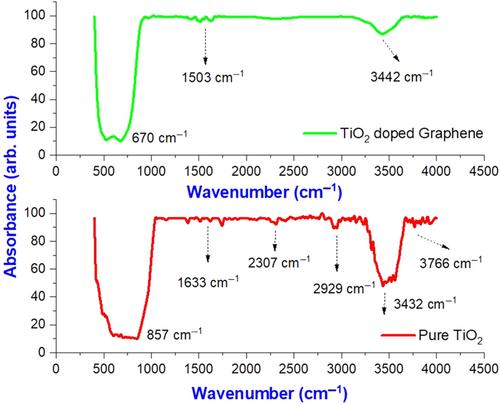
摘要
二氧化钛(TiO2)纳米粒子是通过氧化石墨烯(GO)与茵陈叶提取物反应制成的。X 射线衍射(XRD)分析表明,掺杂了 GO 的二氧化钛纳米颗粒由多种锐钛矿相组成。最初,为了确认掺杂 TiO2 的 GO 纳米粒子(NP's)的生物生成,进行了紫外可见光谱分析。通过扫描电子显微镜,研究表明生物合成的 TiO2 纳米粒子大多呈球形、多分散、纳米级大小。能量色散 X 射线光谱(EDS)图显示,钛(Ti)和氧(O)的峰值明显而稳定,证明了 TiO2 纳米粒子的形成。傅立叶变换红外光谱(FTIR)表明,萜类化合物、黄酮类化合物和蛋白质参与了掺杂 TiO2 的 GO 纳米粒子的生物合成和生产。通过 2,2-二苯基吡啶肼(DPPH)检测评估了掺杂 TiO2 的 GO 纳米粒子的自由基清除活性。此外,与 TiO2 基质相比,掺杂 TiO2 的 GO 纳米粒子具有更强的抗氧化活性。研究了一系列纯 TiO2 和掺杂 TiO2 的 GO 纳米粒子(5、10、50 和 100 mg/mL)溶液的抗菌活性。在目前的研究中,暴露于纯 TiO2 和掺杂 TiO2 的 GO 纳米粒子的斑马鱼胚胎具有毒性,并且根据浓度的不同,存活率较低。在光催化过程中,由于活性物种捕获实验,O2˙ 和 ˙OH 自由基会迅速产生。据估计,120 分钟后,纯 TiO2 纳米粒子和掺杂 GO 的纳米粒子对甲基橙(MO)的降解效率分别为 80%。研究亮点:紫外-可见吸收光谱在 290 nm 处显示出最大吸光峰。扫描电子显微镜(SEM)显示,掺杂纯 TiO2 的 GO NPs 呈团聚状和球形。在斑马鱼胚胎中进行测试时,TiO2 NPs 在高浓度下具有毒性。而 GO 纳米粒子则表现出更好的抗氧化活性。纳米粒子的抗氧化活性与浓度有关。
Investigate the biological activities of Lawsonia inermis extract synthesized from TiO2 doped graphene oxide nanoparticles
Nanoparticles of titanium dioxide (TiO2) were made by reacting graphene oxide (GO) with Lawsonia inermis leaf extract. X-ray diffraction (XRD) analysis revealed crystalline TiO2 doped GO nanoparticles composed of a variety of anatase phases. Initially, UV–vis spectroscopy was performed to confirm the biogenesis of TiO2 doped GO nanoparticles (NP's). Using SEM, the research showed that the biosynthesized TiO2 nanoparticles were mostly spherical, polydispersed, and of a nanoscale size. Because of the energy dispersive X-ray spectroscopy (EDS) pattern, distinct and robust peaks of titanium (Ti) and oxygen (O) were observed, which were supportive of the formation of TiO2 nanoparticles. By using fourier transform infrared (FTIR) spectroscopy, it was demonstrated that terpenoids, flavonoids, and proteins are involved in the biosynthesis and production of TiO2 doped GO nanoparticles. 2,2-diphenylpicrylhydrazyl (DPPH) assays were conducted to evaluate the free radical scavenging activity of TiO2 doped GO nanoparticles. Additionally, the TiO2 doped GO NPs had enhanced antioxidant activity when compared with the TiO2 matrix. A series of pure TiO2 and TiO2 doped GO nanoparticles (5, 10, 50, and 100 mg/mL) solutions were investigated for their antibacterial activities. In the current study, zebrafish embryos exposed to pure TiO2 and TiO2 doped GO nanoparticles were toxic and suffered a low survival rate based on concentration. During photocatalysis, O2˙ and ˙OH radicals are rapidly produced because of the reactive species trapping experiment. It was estimated that pure TiO2 nanoparticles and those doped with GO were 80% effective in degrading methyl orange(MO) after 120 min, respectively.
Research Highlights
The UV–vis absorption spectra showed a maximum absorbance peak at 290 nm.
SEM, the pure TiO2 doped GO NPs exhibit agglomeration and spherical shape.
When tested in zebrafish embryos, TiO2 NPs are toxic at high concentrations.
GO nanoparticles showed better antioxidant activity.
期刊介绍:
Microscopy Research and Technique (MRT) publishes articles on all aspects of advanced microscopy original architecture and methodologies with applications in the biological, clinical, chemical, and materials sciences. Original basic and applied research as well as technical papers dealing with the various subsets of microscopy are encouraged. MRT is the right form for those developing new microscopy methods or using the microscope to answer key questions in basic and applied research.

 求助内容:
求助内容: 应助结果提醒方式:


