{"title":"基于互补标签学习的病理图像细胞类型分类器对滤泡淋巴瘤分级标准的研究","authors":"Ryoichi Koga , Shingo Koide , Hiromu Tanaka , Kei Taguchi , Mauricio Kugler , Tatsuya Yokota , Koichi Ohshima , Hiroaki Miyoshi , Miharu Nagaishi , Noriaki Hashimoto , Ichiro Takeuchi , Hidekata Hontani","doi":"10.1016/j.micron.2024.103663","DOIUrl":null,"url":null,"abstract":"<div><p>We propose a criterion for grading follicular lymphoma that is consistent with the intuitive evaluation, which is conducted by experienced pathologists. A criterion for grading follicular lymphoma is defined by the World Health Organization (WHO) based on the number of centroblasts and centrocytes within the field of view. However, the WHO criterion is not often used in clinical practice because it is impractical for pathologists to visually identify the cell type of each cell and count the number of centroblasts and centrocytes. Hence, based on the widespread use of digital pathology, we make it practical to identify and count the cell type by using image processing and then construct a criterion for grading based on the number of cells. Here, the problem is that labeling the cell type is not easy even for experienced pathologists. To alleviate this problem, we build a new dataset for cell type classification, which contains the pathologists’ confusion records during labeling, and we construct the cell type classifier using complementary-label learning from this dataset. Then we propose a criterion based on the composition ratio of cell types that is consistent with the pathologists’ grading. Our experiments demonstrate that the classifier can accurately identify cell types and the proposed criterion is more consistent with the pathologists’ grading than the current WHO criterion.</p></div>","PeriodicalId":18501,"journal":{"name":"Micron","volume":null,"pages":null},"PeriodicalIF":2.5000,"publicationDate":"2024-05-30","publicationTypes":"Journal Article","fieldsOfStudy":null,"isOpenAccess":false,"openAccessPdf":"https://www.sciencedirect.com/science/article/pii/S0968432824000805/pdfft?md5=d4ffbeee24762fd5b6ba400d6325268a&pid=1-s2.0-S0968432824000805-main.pdf","citationCount":"0","resultStr":"{\"title\":\"A study of criteria for grading follicular lymphoma using a cell type classifier from pathology images based on complementary-label learning\",\"authors\":\"Ryoichi Koga , Shingo Koide , Hiromu Tanaka , Kei Taguchi , Mauricio Kugler , Tatsuya Yokota , Koichi Ohshima , Hiroaki Miyoshi , Miharu Nagaishi , Noriaki Hashimoto , Ichiro Takeuchi , Hidekata Hontani\",\"doi\":\"10.1016/j.micron.2024.103663\",\"DOIUrl\":null,\"url\":null,\"abstract\":\"<div><p>We propose a criterion for grading follicular lymphoma that is consistent with the intuitive evaluation, which is conducted by experienced pathologists. A criterion for grading follicular lymphoma is defined by the World Health Organization (WHO) based on the number of centroblasts and centrocytes within the field of view. However, the WHO criterion is not often used in clinical practice because it is impractical for pathologists to visually identify the cell type of each cell and count the number of centroblasts and centrocytes. Hence, based on the widespread use of digital pathology, we make it practical to identify and count the cell type by using image processing and then construct a criterion for grading based on the number of cells. Here, the problem is that labeling the cell type is not easy even for experienced pathologists. To alleviate this problem, we build a new dataset for cell type classification, which contains the pathologists’ confusion records during labeling, and we construct the cell type classifier using complementary-label learning from this dataset. Then we propose a criterion based on the composition ratio of cell types that is consistent with the pathologists’ grading. Our experiments demonstrate that the classifier can accurately identify cell types and the proposed criterion is more consistent with the pathologists’ grading than the current WHO criterion.</p></div>\",\"PeriodicalId\":18501,\"journal\":{\"name\":\"Micron\",\"volume\":null,\"pages\":null},\"PeriodicalIF\":2.5000,\"publicationDate\":\"2024-05-30\",\"publicationTypes\":\"Journal Article\",\"fieldsOfStudy\":null,\"isOpenAccess\":false,\"openAccessPdf\":\"https://www.sciencedirect.com/science/article/pii/S0968432824000805/pdfft?md5=d4ffbeee24762fd5b6ba400d6325268a&pid=1-s2.0-S0968432824000805-main.pdf\",\"citationCount\":\"0\",\"resultStr\":null,\"platform\":\"Semanticscholar\",\"paperid\":null,\"PeriodicalName\":\"Micron\",\"FirstCategoryId\":\"5\",\"ListUrlMain\":\"https://www.sciencedirect.com/science/article/pii/S0968432824000805\",\"RegionNum\":3,\"RegionCategory\":\"工程技术\",\"ArticlePicture\":[],\"TitleCN\":null,\"AbstractTextCN\":null,\"PMCID\":null,\"EPubDate\":\"\",\"PubModel\":\"\",\"JCR\":\"Q1\",\"JCRName\":\"MICROSCOPY\",\"Score\":null,\"Total\":0}","platform":"Semanticscholar","paperid":null,"PeriodicalName":"Micron","FirstCategoryId":"5","ListUrlMain":"https://www.sciencedirect.com/science/article/pii/S0968432824000805","RegionNum":3,"RegionCategory":"工程技术","ArticlePicture":[],"TitleCN":null,"AbstractTextCN":null,"PMCID":null,"EPubDate":"","PubModel":"","JCR":"Q1","JCRName":"MICROSCOPY","Score":null,"Total":0}
A study of criteria for grading follicular lymphoma using a cell type classifier from pathology images based on complementary-label learning
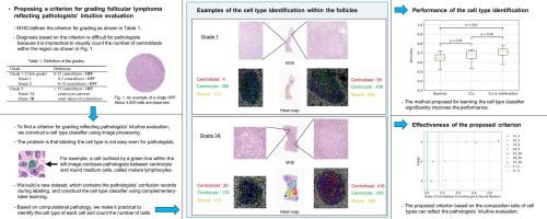
We propose a criterion for grading follicular lymphoma that is consistent with the intuitive evaluation, which is conducted by experienced pathologists. A criterion for grading follicular lymphoma is defined by the World Health Organization (WHO) based on the number of centroblasts and centrocytes within the field of view. However, the WHO criterion is not often used in clinical practice because it is impractical for pathologists to visually identify the cell type of each cell and count the number of centroblasts and centrocytes. Hence, based on the widespread use of digital pathology, we make it practical to identify and count the cell type by using image processing and then construct a criterion for grading based on the number of cells. Here, the problem is that labeling the cell type is not easy even for experienced pathologists. To alleviate this problem, we build a new dataset for cell type classification, which contains the pathologists’ confusion records during labeling, and we construct the cell type classifier using complementary-label learning from this dataset. Then we propose a criterion based on the composition ratio of cell types that is consistent with the pathologists’ grading. Our experiments demonstrate that the classifier can accurately identify cell types and the proposed criterion is more consistent with the pathologists’ grading than the current WHO criterion.
期刊介绍:
Micron is an interdisciplinary forum for all work that involves new applications of microscopy or where advanced microscopy plays a central role. The journal will publish on the design, methods, application, practice or theory of microscopy and microanalysis, including reports on optical, electron-beam, X-ray microtomography, and scanning-probe systems. It also aims at the regular publication of review papers, short communications, as well as thematic issues on contemporary developments in microscopy and microanalysis. The journal embraces original research in which microscopy has contributed significantly to knowledge in biology, life science, nanoscience and nanotechnology, materials science and engineering.
 求助内容:
求助内容: 应助结果提醒方式:


