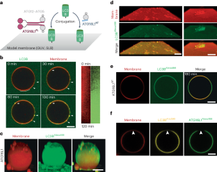
ATG16L1 可诱导形成类似吞噬细胞的膜杯

IF 12.5 1区 生物学 Q1 BIOCHEMISTRY & MOLECULAR BIOLOGY
Jagan Mohan, Satish B. Moparthi, Christine Girard-Blanc, Daniele Campisi, Stéphane Blanchard, Charlotte Nugues, Sowmya Rama, Audrey Salles, Esthel Pénard, Stéphane Vassilopoulos, Thomas Wollert
{"title":"ATG16L1 可诱导形成类似吞噬细胞的膜杯","authors":"Jagan Mohan, Satish B. Moparthi, Christine Girard-Blanc, Daniele Campisi, Stéphane Blanchard, Charlotte Nugues, Sowmya Rama, Audrey Salles, Esthel Pénard, Stéphane Vassilopoulos, Thomas Wollert","doi":"10.1038/s41594-024-01300-y","DOIUrl":null,"url":null,"abstract":"The hallmark of non-selective autophagy is the formation of cup-shaped phagophores that capture bulk cytoplasm. The process is accompanied by the conjugation of LC3B to phagophores by an E3 ligase complex comprising ATG12–ATG5 and ATG16L1. Here we combined two complementary reconstitution approaches to reveal the function of LC3B and its ligase complex during phagophore expansion. We found that LC3B forms together with ATG12–ATG5–ATG16L1 a membrane coat that remodels flat membranes into cups that closely resemble phagophores. Mechanistically, we revealed that cup formation strictly depends on a close collaboration between LC3B and ATG16L1. Moreover, only LC3B, but no other member of the ATG8 protein family, promotes cup formation. ATG16L1 truncates that lacked the C-terminal membrane binding domain catalyzed LC3B lipidation but failed to assemble coats, did not promote cup formation and inhibited the biogenesis of non-selective autophagosomes. Our results thus demonstrate that ATG16L1 and LC3B induce and stabilize the characteristic cup-like shape of phagophores. Autophagy degrades cellular waste by engulfing it in phagophore membranes and delivering it to lysosomes for degradation. Here Mohan and colleagues identified a type of membrane coat that assembles on phagophores to guide their expansion.","PeriodicalId":49141,"journal":{"name":"Nature Structural & Molecular Biology","volume":"31 9","pages":"1448-1459"},"PeriodicalIF":12.5000,"publicationDate":"2024-06-04","publicationTypes":"Journal Article","fieldsOfStudy":null,"isOpenAccess":false,"openAccessPdf":"","citationCount":"0","resultStr":"{\"title\":\"ATG16L1 induces the formation of phagophore-like membrane cups\",\"authors\":\"Jagan Mohan, Satish B. Moparthi, Christine Girard-Blanc, Daniele Campisi, Stéphane Blanchard, Charlotte Nugues, Sowmya Rama, Audrey Salles, Esthel Pénard, Stéphane Vassilopoulos, Thomas Wollert\",\"doi\":\"10.1038/s41594-024-01300-y\",\"DOIUrl\":null,\"url\":null,\"abstract\":\"The hallmark of non-selective autophagy is the formation of cup-shaped phagophores that capture bulk cytoplasm. The process is accompanied by the conjugation of LC3B to phagophores by an E3 ligase complex comprising ATG12–ATG5 and ATG16L1. Here we combined two complementary reconstitution approaches to reveal the function of LC3B and its ligase complex during phagophore expansion. We found that LC3B forms together with ATG12–ATG5–ATG16L1 a membrane coat that remodels flat membranes into cups that closely resemble phagophores. Mechanistically, we revealed that cup formation strictly depends on a close collaboration between LC3B and ATG16L1. Moreover, only LC3B, but no other member of the ATG8 protein family, promotes cup formation. ATG16L1 truncates that lacked the C-terminal membrane binding domain catalyzed LC3B lipidation but failed to assemble coats, did not promote cup formation and inhibited the biogenesis of non-selective autophagosomes. Our results thus demonstrate that ATG16L1 and LC3B induce and stabilize the characteristic cup-like shape of phagophores. Autophagy degrades cellular waste by engulfing it in phagophore membranes and delivering it to lysosomes for degradation. Here Mohan and colleagues identified a type of membrane coat that assembles on phagophores to guide their expansion.\",\"PeriodicalId\":49141,\"journal\":{\"name\":\"Nature Structural & Molecular Biology\",\"volume\":\"31 9\",\"pages\":\"1448-1459\"},\"PeriodicalIF\":12.5000,\"publicationDate\":\"2024-06-04\",\"publicationTypes\":\"Journal Article\",\"fieldsOfStudy\":null,\"isOpenAccess\":false,\"openAccessPdf\":\"\",\"citationCount\":\"0\",\"resultStr\":null,\"platform\":\"Semanticscholar\",\"paperid\":null,\"PeriodicalName\":\"Nature Structural & Molecular Biology\",\"FirstCategoryId\":\"99\",\"ListUrlMain\":\"https://www.nature.com/articles/s41594-024-01300-y\",\"RegionNum\":1,\"RegionCategory\":\"生物学\",\"ArticlePicture\":[],\"TitleCN\":null,\"AbstractTextCN\":null,\"PMCID\":null,\"EPubDate\":\"\",\"PubModel\":\"\",\"JCR\":\"Q1\",\"JCRName\":\"BIOCHEMISTRY & MOLECULAR BIOLOGY\",\"Score\":null,\"Total\":0}","platform":"Semanticscholar","paperid":null,"PeriodicalName":"Nature Structural & Molecular Biology","FirstCategoryId":"99","ListUrlMain":"https://www.nature.com/articles/s41594-024-01300-y","RegionNum":1,"RegionCategory":"生物学","ArticlePicture":[],"TitleCN":null,"AbstractTextCN":null,"PMCID":null,"EPubDate":"","PubModel":"","JCR":"Q1","JCRName":"BIOCHEMISTRY & MOLECULAR BIOLOGY","Score":null,"Total":0}
引用次数: 0

摘要

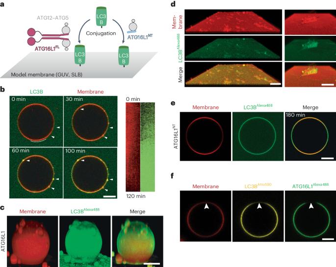
非选择性自噬的特点是形成杯状吞噬体,捕获大量细胞质。在这一过程中,由 ATG12-ATG5 和 ATG16L1 组成的 E3 连接酶复合物会将 LC3B 连接到吞噬体上。在这里,我们结合了两种互补的重组方法,以揭示 LC3B 及其连接酶复合物在吞噬体扩张过程中的功能。我们发现,LC3B与ATG12-ATG5-ATG16L1共同形成了一种膜衣,可将扁平膜重塑为与吞噬体非常相似的杯状膜。从机理上讲,我们发现杯状膜的形成严格依赖于 LC3B 和 ATG16L1 之间的密切合作。此外,只有 LC3B 而非 ATG8 蛋白家族的其他成员能促进杯状结构的形成。缺乏 C 端膜结合结构域的 ATG16L1 截短体能催化 LC3B 脂化,但不能组装外套膜,不能促进杯状形成,并抑制非选择性自噬体的生物生成。因此,我们的研究结果表明,ATG16L1 和 LC3B 能诱导并稳定吞噬体特有的杯状形状。
本文章由计算机程序翻译,如有差异,请以英文原文为准。

ATG16L1 induces the formation of phagophore-like membrane cups

ATG16L1 induces the formation of phagophore-like membrane cups

ATG16L1 induces the formation of phagophore-like membrane cups
The hallmark of non-selective autophagy is the formation of cup-shaped phagophores that capture bulk cytoplasm. The process is accompanied by the conjugation of LC3B to phagophores by an E3 ligase complex comprising ATG12–ATG5 and ATG16L1. Here we combined two complementary reconstitution approaches to reveal the function of LC3B and its ligase complex during phagophore expansion. We found that LC3B forms together with ATG12–ATG5–ATG16L1 a membrane coat that remodels flat membranes into cups that closely resemble phagophores. Mechanistically, we revealed that cup formation strictly depends on a close collaboration between LC3B and ATG16L1. Moreover, only LC3B, but no other member of the ATG8 protein family, promotes cup formation. ATG16L1 truncates that lacked the C-terminal membrane binding domain catalyzed LC3B lipidation but failed to assemble coats, did not promote cup formation and inhibited the biogenesis of non-selective autophagosomes. Our results thus demonstrate that ATG16L1 and LC3B induce and stabilize the characteristic cup-like shape of phagophores. Autophagy degrades cellular waste by engulfing it in phagophore membranes and delivering it to lysosomes for degradation. Here Mohan and colleagues identified a type of membrane coat that assembles on phagophores to guide their expansion.
求助全文
通过发布文献求助,成功后即可免费获取论文全文。 去求助
来源期刊
Nature Structural & Molecular Biology
Nature Structural & Molecular Biology BIOCHEMISTRY & MOLECULAR BIOLOGY-BIOPHYSICS
CiteScore
22.00
自引率
1.80%
发文量
160
审稿时长
3-8 weeks
期刊介绍: Nature Structural & Molecular Biology is a comprehensive platform that combines structural and molecular research. Our journal focuses on exploring the functional and mechanistic aspects of biological processes, emphasizing how molecular components collaborate to achieve a particular function. While structural data can shed light on these insights, our publication does not require them as a prerequisite.
×
引用
GB/T 7714-2015
复制
MLA
复制
APA
复制
导出至
BibTeX EndNote RefMan NoteFirst NoteExpress
×
提示
您的信息不完整,为了账户安全,请先补充。
现在去补充
×
提示
您因"违规操作"
具体请查看互助需知
我知道了
×
提示
确定
请完成安全验证×
copy
已复制链接
快去分享给好友吧!
我知道了
右上角分享
点击右上角分享
0
联系我们:info@booksci.cn Book学术提供免费学术资源搜索服务,方便国内外学者检索中英文文献。致力于提供最便捷和优质的服务体验。 Copyright © 2023 布克学术 All rights reserved.
京ICP备2023020795号-1
ghs 京公网安备 11010802042870号
Book学术文献互助
Book学术文献互助群
群 号:481959085
Book学术官方微信