{"title":"β-谷甾醇通过抑制 GSK3B 的表达减轻肝癌细胞的恶性表型","authors":"Ruoyu Wang, Dan Tang, Longyun Ou, Jiacheng Jiang, Yu-Nan Wu, Xuefei Tian","doi":"10.1007/s13577-024-01081-y","DOIUrl":null,"url":null,"abstract":"<p><p>To explore the effects of β-Sitosterol upon hepatocellular carcinoma cell proliferation, apoptosis, migration, invasion, and epithelial-mesenchymal transition (EMT), and to investigate the underlying mechanism using network pharmacology. Human hepatocellular carcinoma cell lines (Huh-7 and HCCLM3) were expose to gradient concentrations of β-Sitosterol (5 μg/mL, 10 μg/mL, and 20 μg/mL). Cell viability and proliferation were assessed using MTT, CCK-8, colony formation, and EdU assays.Flow cytometry was employed to evaluate cell cycle and apoptosis. Scratch and Transwell assays were performed, respectively, to detect cell migration and invasion. The levels of apoptosis-associated proteins (BAX, BCL2, and cleaved caspase3) as well as EMT-associated proteins (E-cadherin, N-cadherin, Snail, and Vimentin) were detected in Huh-7 and HCCLM3 cell lines using Western blot analysis. The drug target gene for β-Sitosterol was screened via PubChem and subsequently evaluated for expression in the GSE112790 dataset. In addition, the expression level of glycogen synthase kinase 3 beta (GSK3B) within the Cancer Genome Atlas-Liver Hepatocellular Carcinoma (TCGA-LIHC) database was analyzed, along with its correlation to the survival outcomes of patients with hepatocellular carcinoma. The diagnostic efficiency of GSK3B was assessed by analyzing the ROC curve. Subsequently, Huh-7 and HCCLM3 cell lines were transfected with the overexpression vector of GSK3B and then treated with β-Sitosterol to further validate the association between GSK3B and β-Sitosterol. GSK3B demonstrated a significantly elevated expression in patients with hepatocellular carcinoma, which could predict hepatocellular carcinoma patients' impaired prognosis based on GEO dataset and TCGA database. GSK3B inhibitor (CHIR-98014) notably inhibited cell proliferation and invasion, promoted cell apoptosis and cell cycle arrest at G0/G1 phase in hepatocellular carcinoma cells. β-Sitosterol treatment further promoted the efffects of GSK3B inhibitor on hepatocellular carcinoma cells. GSK3B overexpression has been found to enhance the proliferative and invasive capabilities of hepatocellular carcinoma cells. Furthermore it has been observed that GSK3B overexpression, it has been obsear can partially reverse the inhibitory effect of β-Sitosterol upon hepatocellular. β-Sitosterol suppressed hepatocellular carcinoma cell proliferation and invasion, and enhanced apoptosis via inhibiting GSK3B expression.</p>","PeriodicalId":49194,"journal":{"name":"Human Cell","volume":" ","pages":"1156-1169"},"PeriodicalIF":3.4000,"publicationDate":"2024-07-01","publicationTypes":"Journal Article","fieldsOfStudy":null,"isOpenAccess":false,"openAccessPdf":"https://www.ncbi.nlm.nih.gov/pmc/articles/PMC11194219/pdf/","citationCount":"0","resultStr":"{\"title\":\"β-Sitosterol alleviates the malignant phenotype of hepatocellular carcinoma cells via inhibiting GSK3B expression.\",\"authors\":\"Ruoyu Wang, Dan Tang, Longyun Ou, Jiacheng Jiang, Yu-Nan Wu, Xuefei Tian\",\"doi\":\"10.1007/s13577-024-01081-y\",\"DOIUrl\":null,\"url\":null,\"abstract\":\"<p><p>To explore the effects of β-Sitosterol upon hepatocellular carcinoma cell proliferation, apoptosis, migration, invasion, and epithelial-mesenchymal transition (EMT), and to investigate the underlying mechanism using network pharmacology. Human hepatocellular carcinoma cell lines (Huh-7 and HCCLM3) were expose to gradient concentrations of β-Sitosterol (5 μg/mL, 10 μg/mL, and 20 μg/mL). Cell viability and proliferation were assessed using MTT, CCK-8, colony formation, and EdU assays.Flow cytometry was employed to evaluate cell cycle and apoptosis. Scratch and Transwell assays were performed, respectively, to detect cell migration and invasion. The levels of apoptosis-associated proteins (BAX, BCL2, and cleaved caspase3) as well as EMT-associated proteins (E-cadherin, N-cadherin, Snail, and Vimentin) were detected in Huh-7 and HCCLM3 cell lines using Western blot analysis. The drug target gene for β-Sitosterol was screened via PubChem and subsequently evaluated for expression in the GSE112790 dataset. In addition, the expression level of glycogen synthase kinase 3 beta (GSK3B) within the Cancer Genome Atlas-Liver Hepatocellular Carcinoma (TCGA-LIHC) database was analyzed, along with its correlation to the survival outcomes of patients with hepatocellular carcinoma. The diagnostic efficiency of GSK3B was assessed by analyzing the ROC curve. Subsequently, Huh-7 and HCCLM3 cell lines were transfected with the overexpression vector of GSK3B and then treated with β-Sitosterol to further validate the association between GSK3B and β-Sitosterol. GSK3B demonstrated a significantly elevated expression in patients with hepatocellular carcinoma, which could predict hepatocellular carcinoma patients' impaired prognosis based on GEO dataset and TCGA database. GSK3B inhibitor (CHIR-98014) notably inhibited cell proliferation and invasion, promoted cell apoptosis and cell cycle arrest at G0/G1 phase in hepatocellular carcinoma cells. β-Sitosterol treatment further promoted the efffects of GSK3B inhibitor on hepatocellular carcinoma cells. GSK3B overexpression has been found to enhance the proliferative and invasive capabilities of hepatocellular carcinoma cells. Furthermore it has been observed that GSK3B overexpression, it has been obsear can partially reverse the inhibitory effect of β-Sitosterol upon hepatocellular. β-Sitosterol suppressed hepatocellular carcinoma cell proliferation and invasion, and enhanced apoptosis via inhibiting GSK3B expression.</p>\",\"PeriodicalId\":49194,\"journal\":{\"name\":\"Human Cell\",\"volume\":\" \",\"pages\":\"1156-1169\"},\"PeriodicalIF\":3.4000,\"publicationDate\":\"2024-07-01\",\"publicationTypes\":\"Journal Article\",\"fieldsOfStudy\":null,\"isOpenAccess\":false,\"openAccessPdf\":\"https://www.ncbi.nlm.nih.gov/pmc/articles/PMC11194219/pdf/\",\"citationCount\":\"0\",\"resultStr\":null,\"platform\":\"Semanticscholar\",\"paperid\":null,\"PeriodicalName\":\"Human Cell\",\"FirstCategoryId\":\"99\",\"ListUrlMain\":\"https://doi.org/10.1007/s13577-024-01081-y\",\"RegionNum\":3,\"RegionCategory\":\"生物学\",\"ArticlePicture\":[],\"TitleCN\":null,\"AbstractTextCN\":null,\"PMCID\":null,\"EPubDate\":\"2024/5/30 0:00:00\",\"PubModel\":\"Epub\",\"JCR\":\"Q3\",\"JCRName\":\"CELL BIOLOGY\",\"Score\":null,\"Total\":0}","platform":"Semanticscholar","paperid":null,"PeriodicalName":"Human Cell","FirstCategoryId":"99","ListUrlMain":"https://doi.org/10.1007/s13577-024-01081-y","RegionNum":3,"RegionCategory":"生物学","ArticlePicture":[],"TitleCN":null,"AbstractTextCN":null,"PMCID":null,"EPubDate":"2024/5/30 0:00:00","PubModel":"Epub","JCR":"Q3","JCRName":"CELL BIOLOGY","Score":null,"Total":0}
β-Sitosterol alleviates the malignant phenotype of hepatocellular carcinoma cells via inhibiting GSK3B expression.
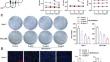
To explore the effects of β-Sitosterol upon hepatocellular carcinoma cell proliferation, apoptosis, migration, invasion, and epithelial-mesenchymal transition (EMT), and to investigate the underlying mechanism using network pharmacology. Human hepatocellular carcinoma cell lines (Huh-7 and HCCLM3) were expose to gradient concentrations of β-Sitosterol (5 μg/mL, 10 μg/mL, and 20 μg/mL). Cell viability and proliferation were assessed using MTT, CCK-8, colony formation, and EdU assays.Flow cytometry was employed to evaluate cell cycle and apoptosis. Scratch and Transwell assays were performed, respectively, to detect cell migration and invasion. The levels of apoptosis-associated proteins (BAX, BCL2, and cleaved caspase3) as well as EMT-associated proteins (E-cadherin, N-cadherin, Snail, and Vimentin) were detected in Huh-7 and HCCLM3 cell lines using Western blot analysis. The drug target gene for β-Sitosterol was screened via PubChem and subsequently evaluated for expression in the GSE112790 dataset. In addition, the expression level of glycogen synthase kinase 3 beta (GSK3B) within the Cancer Genome Atlas-Liver Hepatocellular Carcinoma (TCGA-LIHC) database was analyzed, along with its correlation to the survival outcomes of patients with hepatocellular carcinoma. The diagnostic efficiency of GSK3B was assessed by analyzing the ROC curve. Subsequently, Huh-7 and HCCLM3 cell lines were transfected with the overexpression vector of GSK3B and then treated with β-Sitosterol to further validate the association between GSK3B and β-Sitosterol. GSK3B demonstrated a significantly elevated expression in patients with hepatocellular carcinoma, which could predict hepatocellular carcinoma patients' impaired prognosis based on GEO dataset and TCGA database. GSK3B inhibitor (CHIR-98014) notably inhibited cell proliferation and invasion, promoted cell apoptosis and cell cycle arrest at G0/G1 phase in hepatocellular carcinoma cells. β-Sitosterol treatment further promoted the efffects of GSK3B inhibitor on hepatocellular carcinoma cells. GSK3B overexpression has been found to enhance the proliferative and invasive capabilities of hepatocellular carcinoma cells. Furthermore it has been observed that GSK3B overexpression, it has been obsear can partially reverse the inhibitory effect of β-Sitosterol upon hepatocellular. β-Sitosterol suppressed hepatocellular carcinoma cell proliferation and invasion, and enhanced apoptosis via inhibiting GSK3B expression.
期刊介绍:
Human Cell is the official English-language journal of the Japan Human Cell Society. The journal serves as a forum for international research on all aspects of the human cell, encompassing not only cell biology but also pathology, cytology, and oncology, including clinical oncology. Embryonic stem cells derived from animals, regenerative medicine using animal cells, and experimental animal models with implications for human diseases are covered as well.
Submissions in any of the following categories will be considered: Research Articles, Cell Lines, Rapid Communications, Reviews, and Letters to the Editor. A brief clinical case report focusing on cellular responses to pathological insults in human studies may also be submitted as a Letter to the Editor in a concise and short format.
Not only basic scientists but also gynecologists, oncologists, and other clinical scientists are welcome to submit work expressing new ideas or research using human cells.
 求助内容:
求助内容: 应助结果提醒方式:


