{"title":"通过股骨内侧髁滑动截骨术矫正晚期膝关节骨性关节炎变曲畸形的简易手术技术--手术过程描述和短期疗效--前瞻性研究","authors":"Chandan Pathak, Anjan Chattaraj, Sunit Hazra, Rwivudeep Saha, Sanjay Kumar, Mainak Chandra","doi":"10.1007/s43465-024-01158-8","DOIUrl":null,"url":null,"abstract":"<h3 data-test=\"abstract-sub-heading\">Background</h3><p>Advanced osteoarthritis of knees with varus deformity consists of attenuation of lateral structures with contracture of the posteromedial structures and formation of medial osteophytes. The conventional step-wise medial and posteromedial release with measured resection may sometimes hinder achievement of perfectly balanced flexion and extension gaps with maintenance of flexion stability, without the use of a constrained prosthesis. Medial femoral epicondylar sliding osteotomy tailors the balancing to the need of the kinematics of the native knee and precludes the use of a constrained implant.</p><h3 data-test=\"abstract-sub-heading\">Methods</h3><p>15 patients with Ahlbäck Grades III through V osteoarthritic changes at Howrah Orthopaedic Hospital were included in a prospective cohort case series with a minimum period of follow-up being 12 months. Physical examination, clinical questionnaire and radiographic evaluation were done post-operatively for objectification by functional Knee Society and Oxford Knee Scores respectively.</p><h3 data-test=\"abstract-sub-heading\">Results and Analysis</h3><p>The mean post-operative femorotibial angulation ameliorated to a value of 3.73 ± 1.58° from 18.67 ± 4.2° in the pre-operative stage. The mean overall Range of Motion of operated knee was 109.87 ± 6.86° with no residual frontal laxity and/or laxity in the coronal plane. The mean amount of resection of tibial plateau in patients with severe varus deformity was kept to a minimum, 6.56 mm from the least deficient portion of the lateral condyle. There were no complications as regards component loosening and/or surgical-site infection.</p><h3 data-test=\"abstract-sub-heading\">Conclusion</h3><p>The main objective of balancing a severely varus-afflicted knee is to preserve as much of the Medial Collateral Ligament as possible, to retain its check rein function and not jeopardise the stability. This is ensured by distalisation and posteriorizing the medial epicondyle by an incomplete osteotomy in addition to modest tibial resection fetching a non-isometric point of knee flexion. All osteotomies united by bony union and restoration of kinematic alignment. The limitation of this study however includes the lack of long-term results, such as late instability and polyethylene wear.</p>","PeriodicalId":13338,"journal":{"name":"Indian Journal of Orthopaedics","volume":null,"pages":null},"PeriodicalIF":1.1000,"publicationDate":"2024-05-29","publicationTypes":"Journal Article","fieldsOfStudy":null,"isOpenAccess":false,"openAccessPdf":"","citationCount":"0","resultStr":"{\"title\":\"A Simple Surgical Technique for Correction of Varus Deformity in Advanced Osteoarthritis of Knees by Medial Femoral Condylar Sliding Osteotomy-Description of Procedure and short term Outcome-A Prospective Study\",\"authors\":\"Chandan Pathak, Anjan Chattaraj, Sunit Hazra, Rwivudeep Saha, Sanjay Kumar, Mainak Chandra\",\"doi\":\"10.1007/s43465-024-01158-8\",\"DOIUrl\":null,\"url\":null,\"abstract\":\"<h3 data-test=\\\"abstract-sub-heading\\\">Background</h3><p>Advanced osteoarthritis of knees with varus deformity consists of attenuation of lateral structures with contracture of the posteromedial structures and formation of medial osteophytes. The conventional step-wise medial and posteromedial release with measured resection may sometimes hinder achievement of perfectly balanced flexion and extension gaps with maintenance of flexion stability, without the use of a constrained prosthesis. Medial femoral epicondylar sliding osteotomy tailors the balancing to the need of the kinematics of the native knee and precludes the use of a constrained implant.</p><h3 data-test=\\\"abstract-sub-heading\\\">Methods</h3><p>15 patients with Ahlbäck Grades III through V osteoarthritic changes at Howrah Orthopaedic Hospital were included in a prospective cohort case series with a minimum period of follow-up being 12 months. Physical examination, clinical questionnaire and radiographic evaluation were done post-operatively for objectification by functional Knee Society and Oxford Knee Scores respectively.</p><h3 data-test=\\\"abstract-sub-heading\\\">Results and Analysis</h3><p>The mean post-operative femorotibial angulation ameliorated to a value of 3.73 ± 1.58° from 18.67 ± 4.2° in the pre-operative stage. The mean overall Range of Motion of operated knee was 109.87 ± 6.86° with no residual frontal laxity and/or laxity in the coronal plane. The mean amount of resection of tibial plateau in patients with severe varus deformity was kept to a minimum, 6.56 mm from the least deficient portion of the lateral condyle. There were no complications as regards component loosening and/or surgical-site infection.</p><h3 data-test=\\\"abstract-sub-heading\\\">Conclusion</h3><p>The main objective of balancing a severely varus-afflicted knee is to preserve as much of the Medial Collateral Ligament as possible, to retain its check rein function and not jeopardise the stability. This is ensured by distalisation and posteriorizing the medial epicondyle by an incomplete osteotomy in addition to modest tibial resection fetching a non-isometric point of knee flexion. All osteotomies united by bony union and restoration of kinematic alignment. The limitation of this study however includes the lack of long-term results, such as late instability and polyethylene wear.</p>\",\"PeriodicalId\":13338,\"journal\":{\"name\":\"Indian Journal of Orthopaedics\",\"volume\":null,\"pages\":null},\"PeriodicalIF\":1.1000,\"publicationDate\":\"2024-05-29\",\"publicationTypes\":\"Journal Article\",\"fieldsOfStudy\":null,\"isOpenAccess\":false,\"openAccessPdf\":\"\",\"citationCount\":\"0\",\"resultStr\":null,\"platform\":\"Semanticscholar\",\"paperid\":null,\"PeriodicalName\":\"Indian Journal of Orthopaedics\",\"FirstCategoryId\":\"3\",\"ListUrlMain\":\"https://doi.org/10.1007/s43465-024-01158-8\",\"RegionNum\":4,\"RegionCategory\":\"医学\",\"ArticlePicture\":[],\"TitleCN\":null,\"AbstractTextCN\":null,\"PMCID\":null,\"EPubDate\":\"\",\"PubModel\":\"\",\"JCR\":\"Q3\",\"JCRName\":\"ORTHOPEDICS\",\"Score\":null,\"Total\":0}","platform":"Semanticscholar","paperid":null,"PeriodicalName":"Indian Journal of Orthopaedics","FirstCategoryId":"3","ListUrlMain":"https://doi.org/10.1007/s43465-024-01158-8","RegionNum":4,"RegionCategory":"医学","ArticlePicture":[],"TitleCN":null,"AbstractTextCN":null,"PMCID":null,"EPubDate":"","PubModel":"","JCR":"Q3","JCRName":"ORTHOPEDICS","Score":null,"Total":0}
A Simple Surgical Technique for Correction of Varus Deformity in Advanced Osteoarthritis of Knees by Medial Femoral Condylar Sliding Osteotomy-Description of Procedure and short term Outcome-A Prospective Study
Background
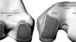
Advanced osteoarthritis of knees with varus deformity consists of attenuation of lateral structures with contracture of the posteromedial structures and formation of medial osteophytes. The conventional step-wise medial and posteromedial release with measured resection may sometimes hinder achievement of perfectly balanced flexion and extension gaps with maintenance of flexion stability, without the use of a constrained prosthesis. Medial femoral epicondylar sliding osteotomy tailors the balancing to the need of the kinematics of the native knee and precludes the use of a constrained implant.
Methods
15 patients with Ahlbäck Grades III through V osteoarthritic changes at Howrah Orthopaedic Hospital were included in a prospective cohort case series with a minimum period of follow-up being 12 months. Physical examination, clinical questionnaire and radiographic evaluation were done post-operatively for objectification by functional Knee Society and Oxford Knee Scores respectively.
Results and Analysis
The mean post-operative femorotibial angulation ameliorated to a value of 3.73 ± 1.58° from 18.67 ± 4.2° in the pre-operative stage. The mean overall Range of Motion of operated knee was 109.87 ± 6.86° with no residual frontal laxity and/or laxity in the coronal plane. The mean amount of resection of tibial plateau in patients with severe varus deformity was kept to a minimum, 6.56 mm from the least deficient portion of the lateral condyle. There were no complications as regards component loosening and/or surgical-site infection.
Conclusion
The main objective of balancing a severely varus-afflicted knee is to preserve as much of the Medial Collateral Ligament as possible, to retain its check rein function and not jeopardise the stability. This is ensured by distalisation and posteriorizing the medial epicondyle by an incomplete osteotomy in addition to modest tibial resection fetching a non-isometric point of knee flexion. All osteotomies united by bony union and restoration of kinematic alignment. The limitation of this study however includes the lack of long-term results, such as late instability and polyethylene wear.
期刊介绍:
IJO welcomes articles that contribute to Orthopaedic knowledge from India and overseas. We publish articles dealing with clinical orthopaedics and basic research in orthopaedic surgery. Articles are accepted only for exclusive publication in the Indian Journal of Orthopaedics. Previously published articles, articles which are in peer-reviewed electronic publications in other journals, are not accepted by the Journal. Published articles and illustrations become the property of the Journal. The copyright remains with the journal. Studies must be carried out in accordance with World Medical Association Declaration of Helsinki.

 求助内容:
求助内容: 应助结果提醒方式:


