{"title":"经褪黑素诱导外泌体(Mel-prExo)处理的人类角膜缘间充质干细胞(hLMSCs)中miR-29、α-SMA和TGFβ1/β3之间的相互关系:洞察角膜无瘢痕愈合。","authors":"Burcugul Altug, Merve Nur Soykan, Sevinc Eyubova, Ayla Eker Sariboyaci, Cezmi Dogan, Onur Ozalp, Eray Atalay","doi":"10.1002/biof.2085","DOIUrl":null,"url":null,"abstract":"<p>Inflammatory mediators that infiltrate the corneal stroma after corneal infections, trauma or refractive surgery can trigger the transformation of corneal keratocytes into myofibroblasts, resulting in highly irregular collagen deposition and subsequently corneal scarring. Mesenchymal stem cells (MSCs) can be used as therapeutic agents to regenerate corneal and conjunctival tissue damage, regulate inflammation, and reduce the development of limbal stem cell failure. The use of MSC-derived exosomes as a cell-free therapeutic vector is a novel therapeutic approach. This study aimed to assess the effect of exosomes obtained from melatonin (Mel)-treated human limbal mesenchymal stem cells (hLMSCs) on naïve hLMSCs and to determine their influence on the antifibrotic and pro-regenerative pathways involved in corneal scarring. hLMSCs were treated with varying concentrations of Mel, followed by isolation and characterization of the procured exosomes (Mel-prExos). These exosomes were added to the cell culture media of naïve hLMSCs to examine their antifibrotic and pro-regenerative effects. The expression of miR-155, miR-29, TGFβ1, TGFβ3, PPARγ, and α-SMA miRNAs and genes were compared between Mel-treated hLMSCs and Mel-prExo-treated hLMSCs by using real-time PCR. We found that at 1 μM Mel and in the presence of Mel-prExos, TGFβ1 was expressed 0.001-fold, while TGFβ3 was expressed 0.6-fold. miR-29 expression was increased 38-fold in the control-Exo group compared to that in the control group. Changes in TGFβ1/β3 and α-SMA expression are associated with miR-29 and miR-155. This approach could prove beneficial for ocular surface tissue engineering applications.</p>","PeriodicalId":8923,"journal":{"name":"BioFactors","volume":"50 6","pages":"1287-1297"},"PeriodicalIF":5.0000,"publicationDate":"2024-05-28","publicationTypes":"Journal Article","fieldsOfStudy":null,"isOpenAccess":false,"openAccessPdf":"https://www.ncbi.nlm.nih.gov/pmc/articles/PMC11627467/pdf/","citationCount":"0","resultStr":"{\"title\":\"Crosstalk among miR-29, α-SMA, and TGFβ1/β3 in melatonin-induced exosome (Mel-prExo) treated human limbal mesenchymal stem cells (hLMSCs): An insight into scarless healing of the cornea\",\"authors\":\"Burcugul Altug, Merve Nur Soykan, Sevinc Eyubova, Ayla Eker Sariboyaci, Cezmi Dogan, Onur Ozalp, Eray Atalay\",\"doi\":\"10.1002/biof.2085\",\"DOIUrl\":null,\"url\":null,\"abstract\":\"<p>Inflammatory mediators that infiltrate the corneal stroma after corneal infections, trauma or refractive surgery can trigger the transformation of corneal keratocytes into myofibroblasts, resulting in highly irregular collagen deposition and subsequently corneal scarring. Mesenchymal stem cells (MSCs) can be used as therapeutic agents to regenerate corneal and conjunctival tissue damage, regulate inflammation, and reduce the development of limbal stem cell failure. The use of MSC-derived exosomes as a cell-free therapeutic vector is a novel therapeutic approach. This study aimed to assess the effect of exosomes obtained from melatonin (Mel)-treated human limbal mesenchymal stem cells (hLMSCs) on naïve hLMSCs and to determine their influence on the antifibrotic and pro-regenerative pathways involved in corneal scarring. hLMSCs were treated with varying concentrations of Mel, followed by isolation and characterization of the procured exosomes (Mel-prExos). These exosomes were added to the cell culture media of naïve hLMSCs to examine their antifibrotic and pro-regenerative effects. The expression of miR-155, miR-29, TGFβ1, TGFβ3, PPARγ, and α-SMA miRNAs and genes were compared between Mel-treated hLMSCs and Mel-prExo-treated hLMSCs by using real-time PCR. We found that at 1 μM Mel and in the presence of Mel-prExos, TGFβ1 was expressed 0.001-fold, while TGFβ3 was expressed 0.6-fold. miR-29 expression was increased 38-fold in the control-Exo group compared to that in the control group. Changes in TGFβ1/β3 and α-SMA expression are associated with miR-29 and miR-155. This approach could prove beneficial for ocular surface tissue engineering applications.</p>\",\"PeriodicalId\":8923,\"journal\":{\"name\":\"BioFactors\",\"volume\":\"50 6\",\"pages\":\"1287-1297\"},\"PeriodicalIF\":5.0000,\"publicationDate\":\"2024-05-28\",\"publicationTypes\":\"Journal Article\",\"fieldsOfStudy\":null,\"isOpenAccess\":false,\"openAccessPdf\":\"https://www.ncbi.nlm.nih.gov/pmc/articles/PMC11627467/pdf/\",\"citationCount\":\"0\",\"resultStr\":null,\"platform\":\"Semanticscholar\",\"paperid\":null,\"PeriodicalName\":\"BioFactors\",\"FirstCategoryId\":\"99\",\"ListUrlMain\":\"https://onlinelibrary.wiley.com/doi/10.1002/biof.2085\",\"RegionNum\":3,\"RegionCategory\":\"生物学\",\"ArticlePicture\":[],\"TitleCN\":null,\"AbstractTextCN\":null,\"PMCID\":null,\"EPubDate\":\"\",\"PubModel\":\"\",\"JCR\":\"Q1\",\"JCRName\":\"BIOCHEMISTRY & MOLECULAR BIOLOGY\",\"Score\":null,\"Total\":0}","platform":"Semanticscholar","paperid":null,"PeriodicalName":"BioFactors","FirstCategoryId":"99","ListUrlMain":"https://onlinelibrary.wiley.com/doi/10.1002/biof.2085","RegionNum":3,"RegionCategory":"生物学","ArticlePicture":[],"TitleCN":null,"AbstractTextCN":null,"PMCID":null,"EPubDate":"","PubModel":"","JCR":"Q1","JCRName":"BIOCHEMISTRY & MOLECULAR BIOLOGY","Score":null,"Total":0}
Crosstalk among miR-29, α-SMA, and TGFβ1/β3 in melatonin-induced exosome (Mel-prExo) treated human limbal mesenchymal stem cells (hLMSCs): An insight into scarless healing of the cornea
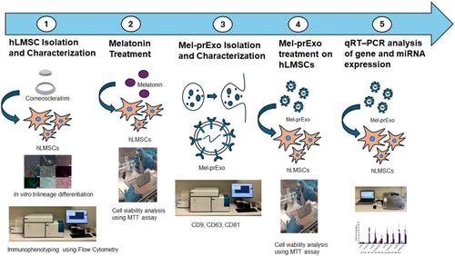
Inflammatory mediators that infiltrate the corneal stroma after corneal infections, trauma or refractive surgery can trigger the transformation of corneal keratocytes into myofibroblasts, resulting in highly irregular collagen deposition and subsequently corneal scarring. Mesenchymal stem cells (MSCs) can be used as therapeutic agents to regenerate corneal and conjunctival tissue damage, regulate inflammation, and reduce the development of limbal stem cell failure. The use of MSC-derived exosomes as a cell-free therapeutic vector is a novel therapeutic approach. This study aimed to assess the effect of exosomes obtained from melatonin (Mel)-treated human limbal mesenchymal stem cells (hLMSCs) on naïve hLMSCs and to determine their influence on the antifibrotic and pro-regenerative pathways involved in corneal scarring. hLMSCs were treated with varying concentrations of Mel, followed by isolation and characterization of the procured exosomes (Mel-prExos). These exosomes were added to the cell culture media of naïve hLMSCs to examine their antifibrotic and pro-regenerative effects. The expression of miR-155, miR-29, TGFβ1, TGFβ3, PPARγ, and α-SMA miRNAs and genes were compared between Mel-treated hLMSCs and Mel-prExo-treated hLMSCs by using real-time PCR. We found that at 1 μM Mel and in the presence of Mel-prExos, TGFβ1 was expressed 0.001-fold, while TGFβ3 was expressed 0.6-fold. miR-29 expression was increased 38-fold in the control-Exo group compared to that in the control group. Changes in TGFβ1/β3 and α-SMA expression are associated with miR-29 and miR-155. This approach could prove beneficial for ocular surface tissue engineering applications.
期刊介绍:
BioFactors, a journal of the International Union of Biochemistry and Molecular Biology, is devoted to the rapid publication of highly significant original research articles and reviews in experimental biology in health and disease.
The word “biofactors” refers to the many compounds that regulate biological functions. Biological factors comprise many molecules produced or modified by living organisms, and present in many essential systems like the blood, the nervous or immunological systems. A non-exhaustive list of biological factors includes neurotransmitters, cytokines, chemokines, hormones, coagulation factors, transcription factors, signaling molecules, receptor ligands and many more. In the group of biofactors we can accommodate several classical molecules not synthetized in the body such as vitamins, micronutrients or essential trace elements.
In keeping with this unified view of biochemistry, BioFactors publishes research dealing with the identification of new substances and the elucidation of their functions at the biophysical, biochemical, cellular and human level as well as studies revealing novel functions of already known biofactors. The journal encourages the submission of studies that use biochemistry, biophysics, cell and molecular biology and/or cell signaling approaches.


 求助内容:
求助内容: 应助结果提醒方式:


