{"title":"羊群发生肠道组织胞浆菌病。","authors":"N Babu Prasath, J Selvaraj","doi":"10.22099/IJVR.2023.47422.6862","DOIUrl":null,"url":null,"abstract":"<p><strong>Background: </strong>The paper describes the first documentary evidence of intestinal histoplasmosis in Indian goats.</p><p><strong>Case description: </strong>A goat flock in delta region of Tamil Nadu with a report of mortality of eight animals with the history of inappetence, constipation, tenesmus and hematochezia. Three goat carcasses were presented for necropsy.</p><p><strong>Findings/treatment and outcome: </strong>Postmortem examination showed mesenteric lymphadenomegaly with greyish yellow viscous contents. Ileum contained scattered dark brownish tubular concretions inside the lumen. Colon and caecum had scattered firm serosal and mucosal nodules with caseous exudate. Lumen contained viscous haemorrhagic content. Mucosa of the colon showed numerous tiny ulcers. Urinary bladder serosa showed a focal firm nodule with caseous exudate and irregular firm mass. Microscopically, lung revealed oval inclusions with clear halo in pulmonary macrophages. Caecum and colon showed ulcers with necrotic core. Necrotic border showed multinucleated giant cells and oval bodies with clear halo. Mesenteric lymph node and urinary bladder revealed pyogranulomatous inflammation with aggregation of macrophages filled with numerous tiny inclusions. Periodic acid-Schiff and Grocott's methanamine silver staining techniques and morphological features of the inclusion confirmed it as <i>Histoplasma capsulatum</i>. The case showed a resemblance with human colonic histoplasmosis.</p><p><strong>Conclusion: </strong>This report documents the first incidence of intestinal histoplasmosis in goats. Interestingly, the intestinal form of histoplasmosis is considered to be common in humans and it showed similarity with human disease in goats based on the gross and microscopic lesions.</p>","PeriodicalId":14629,"journal":{"name":"Iranian journal of veterinary research","volume":"24 4","pages":"357-364"},"PeriodicalIF":1.0000,"publicationDate":"2023-01-01","publicationTypes":"Journal Article","fieldsOfStudy":null,"isOpenAccess":false,"openAccessPdf":"https://www.ncbi.nlm.nih.gov/pmc/articles/PMC11127731/pdf/","citationCount":"0","resultStr":"{\"title\":\"An occurrence of intestinal histoplasmosis in a goat flock.\",\"authors\":\"N Babu Prasath, J Selvaraj\",\"doi\":\"10.22099/IJVR.2023.47422.6862\",\"DOIUrl\":null,\"url\":null,\"abstract\":\"<p><strong>Background: </strong>The paper describes the first documentary evidence of intestinal histoplasmosis in Indian goats.</p><p><strong>Case description: </strong>A goat flock in delta region of Tamil Nadu with a report of mortality of eight animals with the history of inappetence, constipation, tenesmus and hematochezia. Three goat carcasses were presented for necropsy.</p><p><strong>Findings/treatment and outcome: </strong>Postmortem examination showed mesenteric lymphadenomegaly with greyish yellow viscous contents. Ileum contained scattered dark brownish tubular concretions inside the lumen. Colon and caecum had scattered firm serosal and mucosal nodules with caseous exudate. Lumen contained viscous haemorrhagic content. Mucosa of the colon showed numerous tiny ulcers. Urinary bladder serosa showed a focal firm nodule with caseous exudate and irregular firm mass. Microscopically, lung revealed oval inclusions with clear halo in pulmonary macrophages. Caecum and colon showed ulcers with necrotic core. Necrotic border showed multinucleated giant cells and oval bodies with clear halo. Mesenteric lymph node and urinary bladder revealed pyogranulomatous inflammation with aggregation of macrophages filled with numerous tiny inclusions. Periodic acid-Schiff and Grocott's methanamine silver staining techniques and morphological features of the inclusion confirmed it as <i>Histoplasma capsulatum</i>. The case showed a resemblance with human colonic histoplasmosis.</p><p><strong>Conclusion: </strong>This report documents the first incidence of intestinal histoplasmosis in goats. Interestingly, the intestinal form of histoplasmosis is considered to be common in humans and it showed similarity with human disease in goats based on the gross and microscopic lesions.</p>\",\"PeriodicalId\":14629,\"journal\":{\"name\":\"Iranian journal of veterinary research\",\"volume\":\"24 4\",\"pages\":\"357-364\"},\"PeriodicalIF\":1.0000,\"publicationDate\":\"2023-01-01\",\"publicationTypes\":\"Journal Article\",\"fieldsOfStudy\":null,\"isOpenAccess\":false,\"openAccessPdf\":\"https://www.ncbi.nlm.nih.gov/pmc/articles/PMC11127731/pdf/\",\"citationCount\":\"0\",\"resultStr\":null,\"platform\":\"Semanticscholar\",\"paperid\":null,\"PeriodicalName\":\"Iranian journal of veterinary research\",\"FirstCategoryId\":\"97\",\"ListUrlMain\":\"https://doi.org/10.22099/IJVR.2023.47422.6862\",\"RegionNum\":4,\"RegionCategory\":\"农林科学\",\"ArticlePicture\":[],\"TitleCN\":null,\"AbstractTextCN\":null,\"PMCID\":null,\"EPubDate\":\"\",\"PubModel\":\"\",\"JCR\":\"Q3\",\"JCRName\":\"VETERINARY SCIENCES\",\"Score\":null,\"Total\":0}","platform":"Semanticscholar","paperid":null,"PeriodicalName":"Iranian journal of veterinary research","FirstCategoryId":"97","ListUrlMain":"https://doi.org/10.22099/IJVR.2023.47422.6862","RegionNum":4,"RegionCategory":"农林科学","ArticlePicture":[],"TitleCN":null,"AbstractTextCN":null,"PMCID":null,"EPubDate":"","PubModel":"","JCR":"Q3","JCRName":"VETERINARY SCIENCES","Score":null,"Total":0}
An occurrence of intestinal histoplasmosis in a goat flock.
Background: The paper describes the first documentary evidence of intestinal histoplasmosis in Indian goats.
Case description: A goat flock in delta region of Tamil Nadu with a report of mortality of eight animals with the history of inappetence, constipation, tenesmus and hematochezia. Three goat carcasses were presented for necropsy.
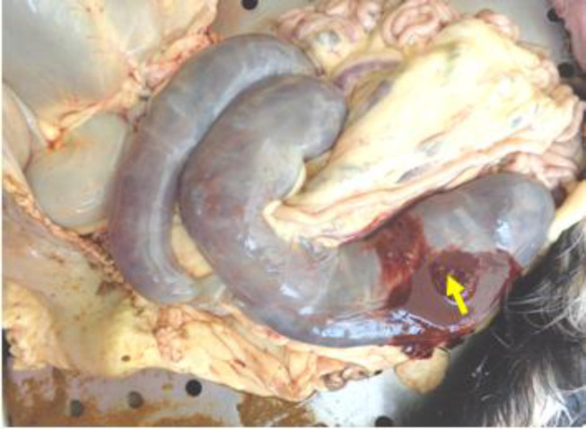
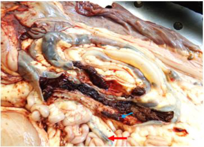
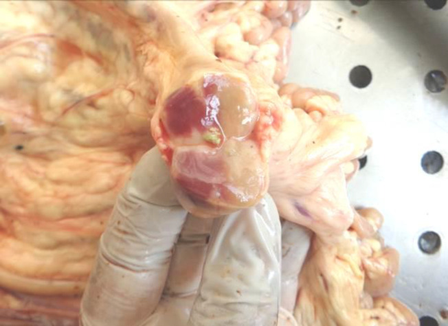
Findings/treatment and outcome: Postmortem examination showed mesenteric lymphadenomegaly with greyish yellow viscous contents. Ileum contained scattered dark brownish tubular concretions inside the lumen. Colon and caecum had scattered firm serosal and mucosal nodules with caseous exudate. Lumen contained viscous haemorrhagic content. Mucosa of the colon showed numerous tiny ulcers. Urinary bladder serosa showed a focal firm nodule with caseous exudate and irregular firm mass. Microscopically, lung revealed oval inclusions with clear halo in pulmonary macrophages. Caecum and colon showed ulcers with necrotic core. Necrotic border showed multinucleated giant cells and oval bodies with clear halo. Mesenteric lymph node and urinary bladder revealed pyogranulomatous inflammation with aggregation of macrophages filled with numerous tiny inclusions. Periodic acid-Schiff and Grocott's methanamine silver staining techniques and morphological features of the inclusion confirmed it as Histoplasma capsulatum. The case showed a resemblance with human colonic histoplasmosis.
Conclusion: This report documents the first incidence of intestinal histoplasmosis in goats. Interestingly, the intestinal form of histoplasmosis is considered to be common in humans and it showed similarity with human disease in goats based on the gross and microscopic lesions.
期刊介绍:
The Iranian Journal of Veterinary Research(IJVR) is published quarterly in 4 issues. The aims of this journal are to improve and expand knowledge in all veterinary fields. It is an international journal indexed by the Thomson Institute for Scientific Information (ISI), Elsevier, Scopus, CAB International, Veterinary Bulletin and several other international databases. Research papers and reports on a wide range of veterinary topics are published in the journal after being evaluated by expert reviewers.The Editor-in-Chief is responsible for the editorial content of the journal—including peer-reviewed manuscripts—and the timing of its publication.
 求助内容:
求助内容: 应助结果提醒方式:


