{"title":"部分水解瓜尔胶通过抑制 NF-κB/MLCK 通路改善败血症患者的肠屏障功能","authors":"Zhaoxia Tang, Yanping Zhu, Xiaoguang Hu, Kayin Lui, Shuhe Li, Xiaodong Song, Changjie Cai, Xiangdong Guan","doi":"10.1007/s12033-024-01180-z","DOIUrl":null,"url":null,"abstract":"<p><p>Partially hydrolyzed guar gum (PHGG) protects against intestinal barrier dysfunction and can ameliorate some intestinal diseases. However, whether PHGG has a role in protecting intestinal barrier function (IBF) during sepsis remains unclear. This study aimed to investigate the role and probable mechanism of PHGG in the intestinal mucosa in sepsis. A rat sepsis model was constructed using cecal ligation and puncture (CLP). FITC-dextran 4 (FD-4) flux, serum inflammatory mediator levels, tight junction (TJ) levels, jejunum mucosa pathology, and epithelial intercellular junction ultrastructure were monitored to evaluate the effect of PHGG on IBF. Caco-2 monolayers were used to study the impact and mechanism of PHGG on lipopolysaccharide (LPS)-induced barrier dysfunction in vitro. The expression of zonula occludens protein-1 and occludin and the location of P65 were studied by immunofluorescence. Nuclear factor kappa B (NF-κB) and myosin light chain kinase 3 (MLCK) pathway-related protein expression was verified by quantitative reverse transcriptase polymerase chain reaction or western blotting. The results indicated that the jejunal mucosa structure was destroyed, the villi were disrupted and shortened, and neutrophil infiltration was evident in the septic rats. Compared to Sham group, spetic rats had increased Chiu's score, serum inflammatory mediator levels, and FD-4 flux but decreased TJ and gap junction density. In addition, the expression of MLCK, p-MLC, and TJ proteins and the expression of P65 in the nucleus were increased in septic rats. Furthermore, compared to those in the Control group, LPS-treated Caco-2 cells showed lower cell viability and transepithelial electrical resistance, while had higher FD-4 flux and the expression of MLCK, p-MLC, TJ proteins and P65 in the nucleus. PHGG pretreatment reversed the above effects induced by CLP or LPS treatment. Moreover, SN50, an NF-κB inhibitor, attenuated the above effects of LPS on Caco-2 cells. Overall, PHGG reduced inflammation, increased TJ protein expression and localization, and relieved damage to the TJ structure and intestinal permeability through suppression of the NF-κB/MLCK pathway. This study provides new insights into the role of PHGG in sepsis therapy.</p>","PeriodicalId":18865,"journal":{"name":"Molecular Biotechnology","volume":" ","pages":"2035-2045"},"PeriodicalIF":2.4000,"publicationDate":"2025-05-01","publicationTypes":"Journal Article","fieldsOfStudy":null,"isOpenAccess":false,"openAccessPdf":"","citationCount":"0","resultStr":"{\"title\":\"Improving Intestinal Barrier Function in Sepsis by Partially Hydrolysed Guar Gum via the Suppression of the NF-κB/MLCK Pathway.\",\"authors\":\"Zhaoxia Tang, Yanping Zhu, Xiaoguang Hu, Kayin Lui, Shuhe Li, Xiaodong Song, Changjie Cai, Xiangdong Guan\",\"doi\":\"10.1007/s12033-024-01180-z\",\"DOIUrl\":null,\"url\":null,\"abstract\":\"<p><p>Partially hydrolyzed guar gum (PHGG) protects against intestinal barrier dysfunction and can ameliorate some intestinal diseases. However, whether PHGG has a role in protecting intestinal barrier function (IBF) during sepsis remains unclear. This study aimed to investigate the role and probable mechanism of PHGG in the intestinal mucosa in sepsis. A rat sepsis model was constructed using cecal ligation and puncture (CLP). FITC-dextran 4 (FD-4) flux, serum inflammatory mediator levels, tight junction (TJ) levels, jejunum mucosa pathology, and epithelial intercellular junction ultrastructure were monitored to evaluate the effect of PHGG on IBF. Caco-2 monolayers were used to study the impact and mechanism of PHGG on lipopolysaccharide (LPS)-induced barrier dysfunction in vitro. The expression of zonula occludens protein-1 and occludin and the location of P65 were studied by immunofluorescence. Nuclear factor kappa B (NF-κB) and myosin light chain kinase 3 (MLCK) pathway-related protein expression was verified by quantitative reverse transcriptase polymerase chain reaction or western blotting. The results indicated that the jejunal mucosa structure was destroyed, the villi were disrupted and shortened, and neutrophil infiltration was evident in the septic rats. Compared to Sham group, spetic rats had increased Chiu's score, serum inflammatory mediator levels, and FD-4 flux but decreased TJ and gap junction density. In addition, the expression of MLCK, p-MLC, and TJ proteins and the expression of P65 in the nucleus were increased in septic rats. Furthermore, compared to those in the Control group, LPS-treated Caco-2 cells showed lower cell viability and transepithelial electrical resistance, while had higher FD-4 flux and the expression of MLCK, p-MLC, TJ proteins and P65 in the nucleus. PHGG pretreatment reversed the above effects induced by CLP or LPS treatment. Moreover, SN50, an NF-κB inhibitor, attenuated the above effects of LPS on Caco-2 cells. Overall, PHGG reduced inflammation, increased TJ protein expression and localization, and relieved damage to the TJ structure and intestinal permeability through suppression of the NF-κB/MLCK pathway. This study provides new insights into the role of PHGG in sepsis therapy.</p>\",\"PeriodicalId\":18865,\"journal\":{\"name\":\"Molecular Biotechnology\",\"volume\":\" \",\"pages\":\"2035-2045\"},\"PeriodicalIF\":2.4000,\"publicationDate\":\"2025-05-01\",\"publicationTypes\":\"Journal Article\",\"fieldsOfStudy\":null,\"isOpenAccess\":false,\"openAccessPdf\":\"\",\"citationCount\":\"0\",\"resultStr\":null,\"platform\":\"Semanticscholar\",\"paperid\":null,\"PeriodicalName\":\"Molecular Biotechnology\",\"FirstCategoryId\":\"3\",\"ListUrlMain\":\"https://doi.org/10.1007/s12033-024-01180-z\",\"RegionNum\":4,\"RegionCategory\":\"生物学\",\"ArticlePicture\":[],\"TitleCN\":null,\"AbstractTextCN\":null,\"PMCID\":null,\"EPubDate\":\"2024/5/24 0:00:00\",\"PubModel\":\"Epub\",\"JCR\":\"Q3\",\"JCRName\":\"BIOCHEMISTRY & MOLECULAR BIOLOGY\",\"Score\":null,\"Total\":0}","platform":"Semanticscholar","paperid":null,"PeriodicalName":"Molecular Biotechnology","FirstCategoryId":"3","ListUrlMain":"https://doi.org/10.1007/s12033-024-01180-z","RegionNum":4,"RegionCategory":"生物学","ArticlePicture":[],"TitleCN":null,"AbstractTextCN":null,"PMCID":null,"EPubDate":"2024/5/24 0:00:00","PubModel":"Epub","JCR":"Q3","JCRName":"BIOCHEMISTRY & MOLECULAR BIOLOGY","Score":null,"Total":0}
Improving Intestinal Barrier Function in Sepsis by Partially Hydrolysed Guar Gum via the Suppression of the NF-κB/MLCK Pathway.
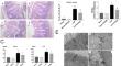
Partially hydrolyzed guar gum (PHGG) protects against intestinal barrier dysfunction and can ameliorate some intestinal diseases. However, whether PHGG has a role in protecting intestinal barrier function (IBF) during sepsis remains unclear. This study aimed to investigate the role and probable mechanism of PHGG in the intestinal mucosa in sepsis. A rat sepsis model was constructed using cecal ligation and puncture (CLP). FITC-dextran 4 (FD-4) flux, serum inflammatory mediator levels, tight junction (TJ) levels, jejunum mucosa pathology, and epithelial intercellular junction ultrastructure were monitored to evaluate the effect of PHGG on IBF. Caco-2 monolayers were used to study the impact and mechanism of PHGG on lipopolysaccharide (LPS)-induced barrier dysfunction in vitro. The expression of zonula occludens protein-1 and occludin and the location of P65 were studied by immunofluorescence. Nuclear factor kappa B (NF-κB) and myosin light chain kinase 3 (MLCK) pathway-related protein expression was verified by quantitative reverse transcriptase polymerase chain reaction or western blotting. The results indicated that the jejunal mucosa structure was destroyed, the villi were disrupted and shortened, and neutrophil infiltration was evident in the septic rats. Compared to Sham group, spetic rats had increased Chiu's score, serum inflammatory mediator levels, and FD-4 flux but decreased TJ and gap junction density. In addition, the expression of MLCK, p-MLC, and TJ proteins and the expression of P65 in the nucleus were increased in septic rats. Furthermore, compared to those in the Control group, LPS-treated Caco-2 cells showed lower cell viability and transepithelial electrical resistance, while had higher FD-4 flux and the expression of MLCK, p-MLC, TJ proteins and P65 in the nucleus. PHGG pretreatment reversed the above effects induced by CLP or LPS treatment. Moreover, SN50, an NF-κB inhibitor, attenuated the above effects of LPS on Caco-2 cells. Overall, PHGG reduced inflammation, increased TJ protein expression and localization, and relieved damage to the TJ structure and intestinal permeability through suppression of the NF-κB/MLCK pathway. This study provides new insights into the role of PHGG in sepsis therapy.
期刊介绍:
Molecular Biotechnology publishes original research papers on the application of molecular biology to both basic and applied research in the field of biotechnology. Particular areas of interest include the following: stability and expression of cloned gene products, cell transformation, gene cloning systems and the production of recombinant proteins, protein purification and analysis, transgenic species, developmental biology, mutation analysis, the applications of DNA fingerprinting, RNA interference, and PCR technology, microarray technology, proteomics, mass spectrometry, bioinformatics, plant molecular biology, microbial genetics, gene probes and the diagnosis of disease, pharmaceutical and health care products, therapeutic agents, vaccines, gene targeting, gene therapy, stem cell technology and tissue engineering, antisense technology, protein engineering and enzyme technology, monoclonal antibodies, glycobiology and glycomics, and agricultural biotechnology.
 求助内容:
求助内容: 应助结果提醒方式:


