{"title":"结直肠癌淋巴结转移中富含亮氨酸重复的 G 蛋白偶联受体 5 的表达:临床病理学见解和预后意义。","authors":"Hiroshi Sawaguchi, Takeshi Uehara, Mai Iwaya, Shiho Asaka, Tomoyuki Nakajima, Masato Kamakura, Tadanobu Nagaya, Takahiro Yoshizawa, Hiroyoshi Ota, Takeji Umemura","doi":"10.1111/pin.13439","DOIUrl":null,"url":null,"abstract":"<p><p>Leucine-rich repeat-containing G protein-coupled receptor 5 (LGR5), a significant cancer stem cell marker in colorectal cancer (CRC), lacks lymph node (LN) expression studies. In this study, we identified LGR5 expression by RNAscope, a highly sensitive RNA in situ method, and analyzed its association with clinicopathological characteristics. Tissue microarrays were generated from primary tumors (PTs) and LN metastases in paraffin-embedded blocks of 38 CRC surgical resection materials. LGR5 expression by RNAscope was evaluated by dividing the expression levels into negative and positive expression. In all but two cases of LN metastasis, LGR5-positive dots were detected in tumor cells, and there was a wide range of LGR5-positive cells. More LGR5-positive dots were identified in the gland-forming region. Twenty-three cases were classified into a high LGR5-expression group, and 15 cases were classified into a low LGR5-expression group. In the high LGR5-expression group, the histological grade was lower than in the low LGR5-expression group (p = 0.0159), while necrosis was significantly more prevalent (p = 0.0326), and the tumor, node, metastasis stage was significantly lower (p = 0.0302). There was no association between LGR5 expression levels in LN metastases and LGR5 expression levels in PT tissue. LGR5 expression in LN metastases may influence prognosis. Further analysis may lead to new therapeutic strategies.</p>","PeriodicalId":19806,"journal":{"name":"Pathology International","volume":" ","pages":"387-393"},"PeriodicalIF":3.4000,"publicationDate":"2024-07-01","publicationTypes":"Journal Article","fieldsOfStudy":null,"isOpenAccess":false,"openAccessPdf":"https://www.ncbi.nlm.nih.gov/pmc/articles/PMC11551821/pdf/","citationCount":"0","resultStr":"{\"title\":\"Leucine-rich repeat-containing G protein-coupled receptor 5 expression in lymph node metastases of colorectal cancer: Clinicopathological insights and prognostic implications.\",\"authors\":\"Hiroshi Sawaguchi, Takeshi Uehara, Mai Iwaya, Shiho Asaka, Tomoyuki Nakajima, Masato Kamakura, Tadanobu Nagaya, Takahiro Yoshizawa, Hiroyoshi Ota, Takeji Umemura\",\"doi\":\"10.1111/pin.13439\",\"DOIUrl\":null,\"url\":null,\"abstract\":\"<p><p>Leucine-rich repeat-containing G protein-coupled receptor 5 (LGR5), a significant cancer stem cell marker in colorectal cancer (CRC), lacks lymph node (LN) expression studies. In this study, we identified LGR5 expression by RNAscope, a highly sensitive RNA in situ method, and analyzed its association with clinicopathological characteristics. Tissue microarrays were generated from primary tumors (PTs) and LN metastases in paraffin-embedded blocks of 38 CRC surgical resection materials. LGR5 expression by RNAscope was evaluated by dividing the expression levels into negative and positive expression. In all but two cases of LN metastasis, LGR5-positive dots were detected in tumor cells, and there was a wide range of LGR5-positive cells. More LGR5-positive dots were identified in the gland-forming region. Twenty-three cases were classified into a high LGR5-expression group, and 15 cases were classified into a low LGR5-expression group. In the high LGR5-expression group, the histological grade was lower than in the low LGR5-expression group (p = 0.0159), while necrosis was significantly more prevalent (p = 0.0326), and the tumor, node, metastasis stage was significantly lower (p = 0.0302). There was no association between LGR5 expression levels in LN metastases and LGR5 expression levels in PT tissue. LGR5 expression in LN metastases may influence prognosis. Further analysis may lead to new therapeutic strategies.</p>\",\"PeriodicalId\":19806,\"journal\":{\"name\":\"Pathology International\",\"volume\":\" \",\"pages\":\"387-393\"},\"PeriodicalIF\":3.4000,\"publicationDate\":\"2024-07-01\",\"publicationTypes\":\"Journal Article\",\"fieldsOfStudy\":null,\"isOpenAccess\":false,\"openAccessPdf\":\"https://www.ncbi.nlm.nih.gov/pmc/articles/PMC11551821/pdf/\",\"citationCount\":\"0\",\"resultStr\":null,\"platform\":\"Semanticscholar\",\"paperid\":null,\"PeriodicalName\":\"Pathology International\",\"FirstCategoryId\":\"3\",\"ListUrlMain\":\"https://doi.org/10.1111/pin.13439\",\"RegionNum\":4,\"RegionCategory\":\"医学\",\"ArticlePicture\":[],\"TitleCN\":null,\"AbstractTextCN\":null,\"PMCID\":null,\"EPubDate\":\"2024/5/24 0:00:00\",\"PubModel\":\"Epub\",\"JCR\":\"Q2\",\"JCRName\":\"PATHOLOGY\",\"Score\":null,\"Total\":0}","platform":"Semanticscholar","paperid":null,"PeriodicalName":"Pathology International","FirstCategoryId":"3","ListUrlMain":"https://doi.org/10.1111/pin.13439","RegionNum":4,"RegionCategory":"医学","ArticlePicture":[],"TitleCN":null,"AbstractTextCN":null,"PMCID":null,"EPubDate":"2024/5/24 0:00:00","PubModel":"Epub","JCR":"Q2","JCRName":"PATHOLOGY","Score":null,"Total":0}
Leucine-rich repeat-containing G protein-coupled receptor 5 expression in lymph node metastases of colorectal cancer: Clinicopathological insights and prognostic implications.
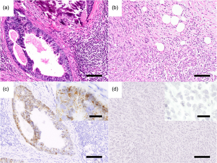
Leucine-rich repeat-containing G protein-coupled receptor 5 (LGR5), a significant cancer stem cell marker in colorectal cancer (CRC), lacks lymph node (LN) expression studies. In this study, we identified LGR5 expression by RNAscope, a highly sensitive RNA in situ method, and analyzed its association with clinicopathological characteristics. Tissue microarrays were generated from primary tumors (PTs) and LN metastases in paraffin-embedded blocks of 38 CRC surgical resection materials. LGR5 expression by RNAscope was evaluated by dividing the expression levels into negative and positive expression. In all but two cases of LN metastasis, LGR5-positive dots were detected in tumor cells, and there was a wide range of LGR5-positive cells. More LGR5-positive dots were identified in the gland-forming region. Twenty-three cases were classified into a high LGR5-expression group, and 15 cases were classified into a low LGR5-expression group. In the high LGR5-expression group, the histological grade was lower than in the low LGR5-expression group (p = 0.0159), while necrosis was significantly more prevalent (p = 0.0326), and the tumor, node, metastasis stage was significantly lower (p = 0.0302). There was no association between LGR5 expression levels in LN metastases and LGR5 expression levels in PT tissue. LGR5 expression in LN metastases may influence prognosis. Further analysis may lead to new therapeutic strategies.
期刊介绍:
Pathology International is the official English journal of the Japanese Society of Pathology, publishing articles of excellence in human and experimental pathology. The Journal focuses on the morphological study of the disease process and/or mechanisms. For human pathology, morphological investigation receives priority but manuscripts describing the result of any ancillary methods (cellular, chemical, immunological and molecular biological) that complement the morphology are accepted. Manuscript on experimental pathology that approach pathologenesis or mechanisms of disease processes are expected to report on the data obtained from models using cellular, biochemical, molecular biological, animal, immunological or other methods in conjunction with morphology. Manuscripts that report data on laboratory medicine (clinical pathology) without significant morphological contribution are not accepted.

 求助内容:
求助内容: 应助结果提醒方式:


