{"title":"通过三维电纺丝制造三维聚己内酯宏观结构。","authors":"Atchara Chinnakorn, Yanawarut Soi-Ngoen, Oratai Weeranantanapan, Phakkhananan Pakawanit, Santi Maensiri, Kriettisak Srisom, Pattanaphong Janphuang, Norbert Radacsi, Wiwat Nuansing","doi":"10.1021/acsbiomaterials.4c00302","DOIUrl":null,"url":null,"abstract":"<p><p>Building 3D electrospun macrostructures and monitoring the biological activities inside them are challenging. In this study, 3D fibrous polycaprolactone (PCL) macrostructures were successfully fabricated using in-house 3D electrospinning. The main factors supporting the 3D self-assembled nanofiber fabrication are the H<sub>3</sub>PO<sub>4</sub> additives, flow rate, and initial distance. The effects of solution concentration, solvent, H<sub>3</sub>PO<sub>4</sub> concentration, flow rate, initial distance, voltage, and nozzle speed on the 3D macrostructures were examined. The optimal conditions of 4 mL/h flow rate, 4 cm initial nozzle-collector distance, 14 kV voltage, and 1 mm/s nozzle speed provided a rapid buildup of cylinder macrostructures with 6 cm of diameter, reaching a final height of 16.18 ± 2.58 mm and a wall thickness of 3.98 ± 1.01 mm on one perimeter with uniform diameter across different sections (1.40 ± 1.10 μm average). Oxygen plasma treatment with 30-50 W for 5 min significantly improved the hydrophilicity of the PCL macrostructures, proving a suitable scaffold for in vitro cell cultures. Additionally, 3D images obtained by synchrotron radiation X-ray tomographic microscopy (SRXTM) presented cell penetration and cell growth within the scaffolds. This breakthrough in 3D electrospinning surpasses current scaffold fabrication limitations, opening new possibilities in various fields.</p>","PeriodicalId":8,"journal":{"name":"ACS Biomaterials Science & Engineering","volume":null,"pages":null},"PeriodicalIF":5.4000,"publicationDate":"2024-05-22","publicationTypes":"Journal Article","fieldsOfStudy":null,"isOpenAccess":false,"openAccessPdf":"","citationCount":"0","resultStr":"{\"title\":\"Fabrication of 3D Polycaprolactone Macrostructures by 3D Electrospinning.\",\"authors\":\"Atchara Chinnakorn, Yanawarut Soi-Ngoen, Oratai Weeranantanapan, Phakkhananan Pakawanit, Santi Maensiri, Kriettisak Srisom, Pattanaphong Janphuang, Norbert Radacsi, Wiwat Nuansing\",\"doi\":\"10.1021/acsbiomaterials.4c00302\",\"DOIUrl\":null,\"url\":null,\"abstract\":\"<p><p>Building 3D electrospun macrostructures and monitoring the biological activities inside them are challenging. In this study, 3D fibrous polycaprolactone (PCL) macrostructures were successfully fabricated using in-house 3D electrospinning. The main factors supporting the 3D self-assembled nanofiber fabrication are the H<sub>3</sub>PO<sub>4</sub> additives, flow rate, and initial distance. The effects of solution concentration, solvent, H<sub>3</sub>PO<sub>4</sub> concentration, flow rate, initial distance, voltage, and nozzle speed on the 3D macrostructures were examined. The optimal conditions of 4 mL/h flow rate, 4 cm initial nozzle-collector distance, 14 kV voltage, and 1 mm/s nozzle speed provided a rapid buildup of cylinder macrostructures with 6 cm of diameter, reaching a final height of 16.18 ± 2.58 mm and a wall thickness of 3.98 ± 1.01 mm on one perimeter with uniform diameter across different sections (1.40 ± 1.10 μm average). Oxygen plasma treatment with 30-50 W for 5 min significantly improved the hydrophilicity of the PCL macrostructures, proving a suitable scaffold for in vitro cell cultures. Additionally, 3D images obtained by synchrotron radiation X-ray tomographic microscopy (SRXTM) presented cell penetration and cell growth within the scaffolds. This breakthrough in 3D electrospinning surpasses current scaffold fabrication limitations, opening new possibilities in various fields.</p>\",\"PeriodicalId\":8,\"journal\":{\"name\":\"ACS Biomaterials Science & Engineering\",\"volume\":null,\"pages\":null},\"PeriodicalIF\":5.4000,\"publicationDate\":\"2024-05-22\",\"publicationTypes\":\"Journal Article\",\"fieldsOfStudy\":null,\"isOpenAccess\":false,\"openAccessPdf\":\"\",\"citationCount\":\"0\",\"resultStr\":null,\"platform\":\"Semanticscholar\",\"paperid\":null,\"PeriodicalName\":\"ACS Biomaterials Science & Engineering\",\"FirstCategoryId\":\"5\",\"ListUrlMain\":\"https://doi.org/10.1021/acsbiomaterials.4c00302\",\"RegionNum\":2,\"RegionCategory\":\"医学\",\"ArticlePicture\":[],\"TitleCN\":null,\"AbstractTextCN\":null,\"PMCID\":null,\"EPubDate\":\"\",\"PubModel\":\"\",\"JCR\":\"Q2\",\"JCRName\":\"MATERIALS SCIENCE, BIOMATERIALS\",\"Score\":null,\"Total\":0}","platform":"Semanticscholar","paperid":null,"PeriodicalName":"ACS Biomaterials Science & Engineering","FirstCategoryId":"5","ListUrlMain":"https://doi.org/10.1021/acsbiomaterials.4c00302","RegionNum":2,"RegionCategory":"医学","ArticlePicture":[],"TitleCN":null,"AbstractTextCN":null,"PMCID":null,"EPubDate":"","PubModel":"","JCR":"Q2","JCRName":"MATERIALS SCIENCE, BIOMATERIALS","Score":null,"Total":0}
Fabrication of 3D Polycaprolactone Macrostructures by 3D Electrospinning.
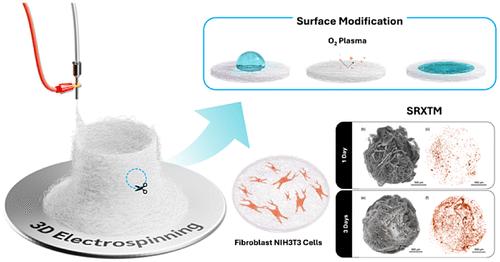
Building 3D electrospun macrostructures and monitoring the biological activities inside them are challenging. In this study, 3D fibrous polycaprolactone (PCL) macrostructures were successfully fabricated using in-house 3D electrospinning. The main factors supporting the 3D self-assembled nanofiber fabrication are the H3PO4 additives, flow rate, and initial distance. The effects of solution concentration, solvent, H3PO4 concentration, flow rate, initial distance, voltage, and nozzle speed on the 3D macrostructures were examined. The optimal conditions of 4 mL/h flow rate, 4 cm initial nozzle-collector distance, 14 kV voltage, and 1 mm/s nozzle speed provided a rapid buildup of cylinder macrostructures with 6 cm of diameter, reaching a final height of 16.18 ± 2.58 mm and a wall thickness of 3.98 ± 1.01 mm on one perimeter with uniform diameter across different sections (1.40 ± 1.10 μm average). Oxygen plasma treatment with 30-50 W for 5 min significantly improved the hydrophilicity of the PCL macrostructures, proving a suitable scaffold for in vitro cell cultures. Additionally, 3D images obtained by synchrotron radiation X-ray tomographic microscopy (SRXTM) presented cell penetration and cell growth within the scaffolds. This breakthrough in 3D electrospinning surpasses current scaffold fabrication limitations, opening new possibilities in various fields.
期刊介绍:
ACS Biomaterials Science & Engineering is the leading journal in the field of biomaterials, serving as an international forum for publishing cutting-edge research and innovative ideas on a broad range of topics:
Applications and Health – implantable tissues and devices, prosthesis, health risks, toxicology
Bio-interactions and Bio-compatibility – material-biology interactions, chemical/morphological/structural communication, mechanobiology, signaling and biological responses, immuno-engineering, calcification, coatings, corrosion and degradation of biomaterials and devices, biophysical regulation of cell functions
Characterization, Synthesis, and Modification – new biomaterials, bioinspired and biomimetic approaches to biomaterials, exploiting structural hierarchy and architectural control, combinatorial strategies for biomaterials discovery, genetic biomaterials design, synthetic biology, new composite systems, bionics, polymer synthesis
Controlled Release and Delivery Systems – biomaterial-based drug and gene delivery, bio-responsive delivery of regulatory molecules, pharmaceutical engineering
Healthcare Advances – clinical translation, regulatory issues, patient safety, emerging trends
Imaging and Diagnostics – imaging agents and probes, theranostics, biosensors, monitoring
Manufacturing and Technology – 3D printing, inks, organ-on-a-chip, bioreactor/perfusion systems, microdevices, BioMEMS, optics and electronics interfaces with biomaterials, systems integration
Modeling and Informatics Tools – scaling methods to guide biomaterial design, predictive algorithms for structure-function, biomechanics, integrating bioinformatics with biomaterials discovery, metabolomics in the context of biomaterials
Tissue Engineering and Regenerative Medicine – basic and applied studies, cell therapies, scaffolds, vascularization, bioartificial organs, transplantation and functionality, cellular agriculture

 求助内容:
求助内容: 应助结果提醒方式:


