Blake W Dieckmann, Marcell E Paguaga, Gary W McCollum, John S Penn, Md Imam Uddin
{"title":"NLRP3 炎症体在脉络膜新生血管中单核细胞和小胶质细胞招募中的作用","authors":"Blake W Dieckmann, Marcell E Paguaga, Gary W McCollum, John S Penn, Md Imam Uddin","doi":"10.4049/immunohorizons.2400025","DOIUrl":null,"url":null,"abstract":"<p><p>Although the pathogenesis of choroidal neovascularization (CNV) is largely unknown in age-related macular degeneration (AMD), inflammasomes may contribute to CNV development and progression. To understand the role NLRP3 inflammasomes in CNV, we used Ccr2RFPCx3cr1GFP dual-reporter mice and immunostaining techniques to confirm localization of NLRP3 inflammasomes in the laser-induced CNV (LCNV) lesions. Confocal microscopy was used to image and quantify LCNV volumes. MCC950 was used as NLRP3 inhibitor. ELISA and quantitative RT-PCR were used to confirm the activation of NLRP3 by monitoring the expression of IL-1β protein and mRNA in choroidal tissues from LCNV mice. In addition, NLRP3 (-/-) LCNV mice were used to investigate whether NLRP3 inflammasomes contribute to the development of LCNV lesions. We observed that red fluorescent protein (RFP)-positive monocyte-derived macrophages and GFP-positive microglia-derived macrophages, in addition to other cell types, were localized in LCNV lesions at day 7 post-laser injury. In addition, NLRP3 inflammasomes are associated with LCNV lesions. Inhibition of NLRP3 inflammasomes, using MCC950, caused an increased Ccr2RFP-positive macrophages, Cx3cr1GFP-positive microglia, and other cells, resulting in an increase in total lesion size. NLRP3 (-/-) LCNV mice showed significantly increased lesion size compared with age-matched controls. Inhibition of NLRP3 resulted in decreased IL-1β mRNA and protein expression in the choroidal tissues, suggesting that increased lesion size may not be directly related to IL-1β.</p>","PeriodicalId":94037,"journal":{"name":"ImmunoHorizons","volume":"8 5","pages":"363-370"},"PeriodicalIF":0.0000,"publicationDate":"2024-05-01","publicationTypes":"Journal Article","fieldsOfStudy":null,"isOpenAccess":false,"openAccessPdf":"https://www.ncbi.nlm.nih.gov/pmc/articles/PMC11150128/pdf/","citationCount":"0","resultStr":"{\"title\":\"Role of NLRP3 Inflammasomes in Monocyte and Microglial Recruitments in Choroidal Neovascularization.\",\"authors\":\"Blake W Dieckmann, Marcell E Paguaga, Gary W McCollum, John S Penn, Md Imam Uddin\",\"doi\":\"10.4049/immunohorizons.2400025\",\"DOIUrl\":null,\"url\":null,\"abstract\":\"<p><p>Although the pathogenesis of choroidal neovascularization (CNV) is largely unknown in age-related macular degeneration (AMD), inflammasomes may contribute to CNV development and progression. To understand the role NLRP3 inflammasomes in CNV, we used Ccr2RFPCx3cr1GFP dual-reporter mice and immunostaining techniques to confirm localization of NLRP3 inflammasomes in the laser-induced CNV (LCNV) lesions. Confocal microscopy was used to image and quantify LCNV volumes. MCC950 was used as NLRP3 inhibitor. ELISA and quantitative RT-PCR were used to confirm the activation of NLRP3 by monitoring the expression of IL-1β protein and mRNA in choroidal tissues from LCNV mice. In addition, NLRP3 (-/-) LCNV mice were used to investigate whether NLRP3 inflammasomes contribute to the development of LCNV lesions. We observed that red fluorescent protein (RFP)-positive monocyte-derived macrophages and GFP-positive microglia-derived macrophages, in addition to other cell types, were localized in LCNV lesions at day 7 post-laser injury. In addition, NLRP3 inflammasomes are associated with LCNV lesions. Inhibition of NLRP3 inflammasomes, using MCC950, caused an increased Ccr2RFP-positive macrophages, Cx3cr1GFP-positive microglia, and other cells, resulting in an increase in total lesion size. NLRP3 (-/-) LCNV mice showed significantly increased lesion size compared with age-matched controls. Inhibition of NLRP3 resulted in decreased IL-1β mRNA and protein expression in the choroidal tissues, suggesting that increased lesion size may not be directly related to IL-1β.</p>\",\"PeriodicalId\":94037,\"journal\":{\"name\":\"ImmunoHorizons\",\"volume\":\"8 5\",\"pages\":\"363-370\"},\"PeriodicalIF\":0.0000,\"publicationDate\":\"2024-05-01\",\"publicationTypes\":\"Journal Article\",\"fieldsOfStudy\":null,\"isOpenAccess\":false,\"openAccessPdf\":\"https://www.ncbi.nlm.nih.gov/pmc/articles/PMC11150128/pdf/\",\"citationCount\":\"0\",\"resultStr\":null,\"platform\":\"Semanticscholar\",\"paperid\":null,\"PeriodicalName\":\"ImmunoHorizons\",\"FirstCategoryId\":\"1085\",\"ListUrlMain\":\"https://doi.org/10.4049/immunohorizons.2400025\",\"RegionNum\":0,\"RegionCategory\":null,\"ArticlePicture\":[],\"TitleCN\":null,\"AbstractTextCN\":null,\"PMCID\":null,\"EPubDate\":\"\",\"PubModel\":\"\",\"JCR\":\"Q3\",\"JCRName\":\"Medicine\",\"Score\":null,\"Total\":0}","platform":"Semanticscholar","paperid":null,"PeriodicalName":"ImmunoHorizons","FirstCategoryId":"1085","ListUrlMain":"https://doi.org/10.4049/immunohorizons.2400025","RegionNum":0,"RegionCategory":null,"ArticlePicture":[],"TitleCN":null,"AbstractTextCN":null,"PMCID":null,"EPubDate":"","PubModel":"","JCR":"Q3","JCRName":"Medicine","Score":null,"Total":0}
Role of NLRP3 Inflammasomes in Monocyte and Microglial Recruitments in Choroidal Neovascularization.
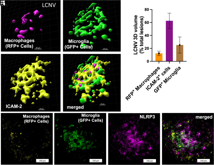
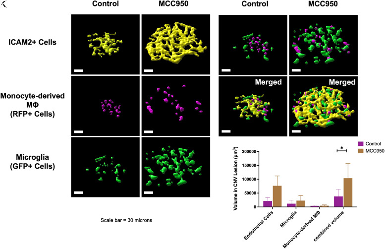
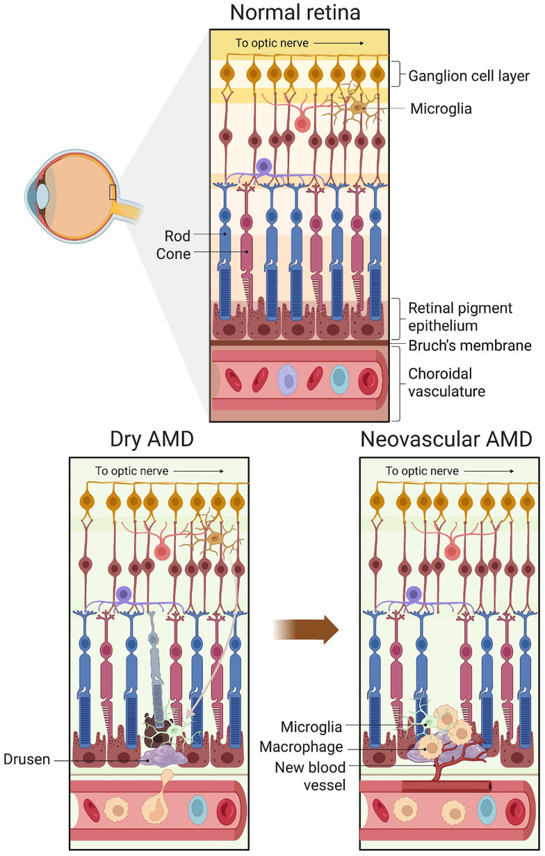
Although the pathogenesis of choroidal neovascularization (CNV) is largely unknown in age-related macular degeneration (AMD), inflammasomes may contribute to CNV development and progression. To understand the role NLRP3 inflammasomes in CNV, we used Ccr2RFPCx3cr1GFP dual-reporter mice and immunostaining techniques to confirm localization of NLRP3 inflammasomes in the laser-induced CNV (LCNV) lesions. Confocal microscopy was used to image and quantify LCNV volumes. MCC950 was used as NLRP3 inhibitor. ELISA and quantitative RT-PCR were used to confirm the activation of NLRP3 by monitoring the expression of IL-1β protein and mRNA in choroidal tissues from LCNV mice. In addition, NLRP3 (-/-) LCNV mice were used to investigate whether NLRP3 inflammasomes contribute to the development of LCNV lesions. We observed that red fluorescent protein (RFP)-positive monocyte-derived macrophages and GFP-positive microglia-derived macrophages, in addition to other cell types, were localized in LCNV lesions at day 7 post-laser injury. In addition, NLRP3 inflammasomes are associated with LCNV lesions. Inhibition of NLRP3 inflammasomes, using MCC950, caused an increased Ccr2RFP-positive macrophages, Cx3cr1GFP-positive microglia, and other cells, resulting in an increase in total lesion size. NLRP3 (-/-) LCNV mice showed significantly increased lesion size compared with age-matched controls. Inhibition of NLRP3 resulted in decreased IL-1β mRNA and protein expression in the choroidal tissues, suggesting that increased lesion size may not be directly related to IL-1β.

 求助内容:
求助内容: 应助结果提醒方式:


