{"title":"髌骨股骨痛患者在负重姿势下腓肠肌的大小和激活能力受损。","authors":"Abbis Jaffri, Amber Schwarting, Andrea Baellow","doi":"10.1002/jfa2.12014","DOIUrl":null,"url":null,"abstract":"<p><strong>Background: </strong>Patellofemoral pain (PFP) is characterized by chronic pain in the anterior aspect of the knee during loading activities. Many studies investigating muscle morphology changes for individuals with PFP focus on the proximal joints, however, few studies have investigated muscles of the foot and ankle complex. This study aimed to explore the differences in peroneal muscle size and activation between individuals with PFP and healthy controls using ultrasound imaging in weight-bearing.</p><p><strong>Methods: </strong>A case-control study in a university lab setting was conducted. Thirty individuals with PFP (age: 20.23 ± 3.30 years, mass: 74.70 ± 27.63 kgs, height: 161.32 ± 11.72 cm) and 30 healthy individuals (age: 20.33 ± 3.37 years, mass: 64.02 ± 11.00 kgs, height: 169.31 ± 9.30 cm) participated. Cross-sectional area (CSA) images of the peroneal muscles were taken in non-weight bearing and weight-bearing positions. The functional activation ratio from lying to single-leg standing (SLS) was calculated.</p><p><strong>Results: </strong>There was a statistically significant (p = 0.041) group (PFP, healthy) by position (non-weight-bearing, weight-bearing) interaction for the peroneal muscle CSA with a Cohen's d effect size of 0.2 in non-weight-bearing position and 0.7 in weight-bearing position. The functional activation ratio for the healthy group was significantly more (p = 0.01) than the PFP group.</p><p><strong>Conclusion: </strong>Peroneal muscles were found to be smaller in size in those with PFP compared to the healthy subjects in the weight-bearing SLS position. This study found that those with PFP have lower activation of peroneal muscles in functional position.</p>","PeriodicalId":49164,"journal":{"name":"Journal of Foot and Ankle Research","volume":"17 2","pages":"e12014"},"PeriodicalIF":2.2000,"publicationDate":"2024-06-01","publicationTypes":"Journal Article","fieldsOfStudy":null,"isOpenAccess":false,"openAccessPdf":"https://www.ncbi.nlm.nih.gov/pmc/articles/PMC11296708/pdf/","citationCount":"0","resultStr":"{\"title\":\"Impairments in peroneal muscle size and activation in individuals with patellofemoral pain in weight-bearing position.\",\"authors\":\"Abbis Jaffri, Amber Schwarting, Andrea Baellow\",\"doi\":\"10.1002/jfa2.12014\",\"DOIUrl\":null,\"url\":null,\"abstract\":\"<p><strong>Background: </strong>Patellofemoral pain (PFP) is characterized by chronic pain in the anterior aspect of the knee during loading activities. Many studies investigating muscle morphology changes for individuals with PFP focus on the proximal joints, however, few studies have investigated muscles of the foot and ankle complex. This study aimed to explore the differences in peroneal muscle size and activation between individuals with PFP and healthy controls using ultrasound imaging in weight-bearing.</p><p><strong>Methods: </strong>A case-control study in a university lab setting was conducted. Thirty individuals with PFP (age: 20.23 ± 3.30 years, mass: 74.70 ± 27.63 kgs, height: 161.32 ± 11.72 cm) and 30 healthy individuals (age: 20.33 ± 3.37 years, mass: 64.02 ± 11.00 kgs, height: 169.31 ± 9.30 cm) participated. Cross-sectional area (CSA) images of the peroneal muscles were taken in non-weight bearing and weight-bearing positions. The functional activation ratio from lying to single-leg standing (SLS) was calculated.</p><p><strong>Results: </strong>There was a statistically significant (p = 0.041) group (PFP, healthy) by position (non-weight-bearing, weight-bearing) interaction for the peroneal muscle CSA with a Cohen's d effect size of 0.2 in non-weight-bearing position and 0.7 in weight-bearing position. The functional activation ratio for the healthy group was significantly more (p = 0.01) than the PFP group.</p><p><strong>Conclusion: </strong>Peroneal muscles were found to be smaller in size in those with PFP compared to the healthy subjects in the weight-bearing SLS position. This study found that those with PFP have lower activation of peroneal muscles in functional position.</p>\",\"PeriodicalId\":49164,\"journal\":{\"name\":\"Journal of Foot and Ankle Research\",\"volume\":\"17 2\",\"pages\":\"e12014\"},\"PeriodicalIF\":2.2000,\"publicationDate\":\"2024-06-01\",\"publicationTypes\":\"Journal Article\",\"fieldsOfStudy\":null,\"isOpenAccess\":false,\"openAccessPdf\":\"https://www.ncbi.nlm.nih.gov/pmc/articles/PMC11296708/pdf/\",\"citationCount\":\"0\",\"resultStr\":null,\"platform\":\"Semanticscholar\",\"paperid\":null,\"PeriodicalName\":\"Journal of Foot and Ankle Research\",\"FirstCategoryId\":\"3\",\"ListUrlMain\":\"https://doi.org/10.1002/jfa2.12014\",\"RegionNum\":3,\"RegionCategory\":\"医学\",\"ArticlePicture\":[],\"TitleCN\":null,\"AbstractTextCN\":null,\"PMCID\":null,\"EPubDate\":\"\",\"PubModel\":\"\",\"JCR\":\"Q1\",\"JCRName\":\"ORTHOPEDICS\",\"Score\":null,\"Total\":0}","platform":"Semanticscholar","paperid":null,"PeriodicalName":"Journal of Foot and Ankle Research","FirstCategoryId":"3","ListUrlMain":"https://doi.org/10.1002/jfa2.12014","RegionNum":3,"RegionCategory":"医学","ArticlePicture":[],"TitleCN":null,"AbstractTextCN":null,"PMCID":null,"EPubDate":"","PubModel":"","JCR":"Q1","JCRName":"ORTHOPEDICS","Score":null,"Total":0}
Impairments in peroneal muscle size and activation in individuals with patellofemoral pain in weight-bearing position.
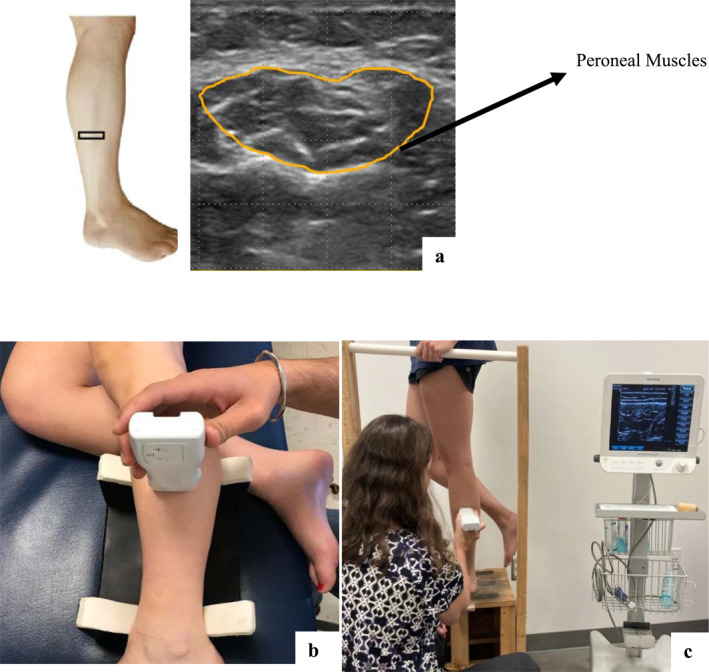
Background: Patellofemoral pain (PFP) is characterized by chronic pain in the anterior aspect of the knee during loading activities. Many studies investigating muscle morphology changes for individuals with PFP focus on the proximal joints, however, few studies have investigated muscles of the foot and ankle complex. This study aimed to explore the differences in peroneal muscle size and activation between individuals with PFP and healthy controls using ultrasound imaging in weight-bearing.
Methods: A case-control study in a university lab setting was conducted. Thirty individuals with PFP (age: 20.23 ± 3.30 years, mass: 74.70 ± 27.63 kgs, height: 161.32 ± 11.72 cm) and 30 healthy individuals (age: 20.33 ± 3.37 years, mass: 64.02 ± 11.00 kgs, height: 169.31 ± 9.30 cm) participated. Cross-sectional area (CSA) images of the peroneal muscles were taken in non-weight bearing and weight-bearing positions. The functional activation ratio from lying to single-leg standing (SLS) was calculated.
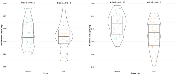
Results: There was a statistically significant (p = 0.041) group (PFP, healthy) by position (non-weight-bearing, weight-bearing) interaction for the peroneal muscle CSA with a Cohen's d effect size of 0.2 in non-weight-bearing position and 0.7 in weight-bearing position. The functional activation ratio for the healthy group was significantly more (p = 0.01) than the PFP group.
Conclusion: Peroneal muscles were found to be smaller in size in those with PFP compared to the healthy subjects in the weight-bearing SLS position. This study found that those with PFP have lower activation of peroneal muscles in functional position.
期刊介绍:
Journal of Foot and Ankle Research, the official journal of the Australian Podiatry Association and The College of Podiatry (UK), is an open access journal that encompasses all aspects of policy, organisation, delivery and clinical practice related to the assessment, diagnosis, prevention and management of foot and ankle disorders.
Journal of Foot and Ankle Research covers a wide range of clinical subject areas, including diabetology, paediatrics, sports medicine, gerontology and geriatrics, foot surgery, physical therapy, dermatology, wound management, radiology, biomechanics and bioengineering, orthotics and prosthetics, as well the broad areas of epidemiology, policy, organisation and delivery of services related to foot and ankle care.
The journal encourages submissions from all health professionals who manage lower limb conditions, including podiatrists, nurses, physical therapists and physiotherapists, orthopaedists, manual therapists, medical specialists and general medical practitioners, as well as health service researchers concerned with foot and ankle care.
The Australian Podiatry Association and the College of Podiatry (UK) have reserve funds to cover the article-processing charge for manuscripts submitted by its members. Society members can email the appropriate contact at Australian Podiatry Association or The College of Podiatry to obtain the corresponding code to enter on submission.
 求助内容:
求助内容: 应助结果提醒方式:


