Ian Towle, Carolina Loch, Marc Oxenham, Kristin L. Krueger, Amira Samir Salem, Marina Martínez de Pinillos, Mario Modesto-Mata, Leslea J. Hlusko
{"title":"技术说明:利用牙科组织进行骨矿物质研究的微型计算机断层扫描校准。","authors":"Ian Towle, Carolina Loch, Marc Oxenham, Kristin L. Krueger, Amira Samir Salem, Marina Martínez de Pinillos, Mario Modesto-Mata, Leslea J. Hlusko","doi":"10.1002/ajpa.24952","DOIUrl":null,"url":null,"abstract":"<p>Computed tomography (CT) and microcomputed tomography (μCT) require calibration against density phantoms scanned with specimens or during routine internal calibration for assessment of mineral concentration (MC) and density. In clinical studies involving bone, alternative calibration methods using bodily tissues and fluids (“phantomless” calibration) have been suggested. However, such tissues are seldom available in archeological and osteological research. This study investigates the potential of dental tissue as internal reference for calibration of μCT scans, facilitating the analysis of bone MC. We analyzed 70 molars from 24 extant primate species, including eight human teeth, each scanned with density phantoms for calibration. Our findings indicate that sampling specific regions of molars (lateral aspects of the mesial cusps) yields low variation in enamel and dentine MC values, averaging 1.27 g/cm<sup>3</sup> (±0.03) for dentine and 2.25 g/cm<sup>3</sup> (±0.03) for enamel. No significant differences were observed across molar types or among scanning procedures, including scanner model, resolution, and filters. An ad hoc test on 12 mandibles revealed low variance in MC between the conventional phantom and dental tissue calibration methods; all 36 measurements (low, medium, and high MC for each mandible) were within 0.05 g/cm<sup>3</sup> of each other —81% were within 0.03 g/cm<sup>3</sup> and 94% within 0.04 g/cm<sup>3</sup>. Based on these results, we propose a new “phantomless” calibration technique using these mean enamel and dentine MC values. The presented phantomless calibration method could aid in the assessment of bone pathology and enhance the scope of studies investigating bone structure and physical property variations in archeological, osteological, and laboratory-based research.</p>","PeriodicalId":29759,"journal":{"name":"American Journal of Biological Anthropology","volume":"184 3","pages":""},"PeriodicalIF":1.7000,"publicationDate":"2024-05-22","publicationTypes":"Journal Article","fieldsOfStudy":null,"isOpenAccess":false,"openAccessPdf":"https://onlinelibrary.wiley.com/doi/epdf/10.1002/ajpa.24952","citationCount":"0","resultStr":"{\"title\":\"Technical note: Micro-computed tomography calibration using dental tissue for bone mineral research\",\"authors\":\"Ian Towle, Carolina Loch, Marc Oxenham, Kristin L. Krueger, Amira Samir Salem, Marina Martínez de Pinillos, Mario Modesto-Mata, Leslea J. Hlusko\",\"doi\":\"10.1002/ajpa.24952\",\"DOIUrl\":null,\"url\":null,\"abstract\":\"<p>Computed tomography (CT) and microcomputed tomography (μCT) require calibration against density phantoms scanned with specimens or during routine internal calibration for assessment of mineral concentration (MC) and density. In clinical studies involving bone, alternative calibration methods using bodily tissues and fluids (“phantomless” calibration) have been suggested. However, such tissues are seldom available in archeological and osteological research. This study investigates the potential of dental tissue as internal reference for calibration of μCT scans, facilitating the analysis of bone MC. We analyzed 70 molars from 24 extant primate species, including eight human teeth, each scanned with density phantoms for calibration. Our findings indicate that sampling specific regions of molars (lateral aspects of the mesial cusps) yields low variation in enamel and dentine MC values, averaging 1.27 g/cm<sup>3</sup> (±0.03) for dentine and 2.25 g/cm<sup>3</sup> (±0.03) for enamel. No significant differences were observed across molar types or among scanning procedures, including scanner model, resolution, and filters. An ad hoc test on 12 mandibles revealed low variance in MC between the conventional phantom and dental tissue calibration methods; all 36 measurements (low, medium, and high MC for each mandible) were within 0.05 g/cm<sup>3</sup> of each other —81% were within 0.03 g/cm<sup>3</sup> and 94% within 0.04 g/cm<sup>3</sup>. Based on these results, we propose a new “phantomless” calibration technique using these mean enamel and dentine MC values. The presented phantomless calibration method could aid in the assessment of bone pathology and enhance the scope of studies investigating bone structure and physical property variations in archeological, osteological, and laboratory-based research.</p>\",\"PeriodicalId\":29759,\"journal\":{\"name\":\"American Journal of Biological Anthropology\",\"volume\":\"184 3\",\"pages\":\"\"},\"PeriodicalIF\":1.7000,\"publicationDate\":\"2024-05-22\",\"publicationTypes\":\"Journal Article\",\"fieldsOfStudy\":null,\"isOpenAccess\":false,\"openAccessPdf\":\"https://onlinelibrary.wiley.com/doi/epdf/10.1002/ajpa.24952\",\"citationCount\":\"0\",\"resultStr\":null,\"platform\":\"Semanticscholar\",\"paperid\":null,\"PeriodicalName\":\"American Journal of Biological Anthropology\",\"FirstCategoryId\":\"1085\",\"ListUrlMain\":\"https://onlinelibrary.wiley.com/doi/10.1002/ajpa.24952\",\"RegionNum\":2,\"RegionCategory\":\"生物学\",\"ArticlePicture\":[],\"TitleCN\":null,\"AbstractTextCN\":null,\"PMCID\":null,\"EPubDate\":\"\",\"PubModel\":\"\",\"JCR\":\"Q1\",\"JCRName\":\"ANTHROPOLOGY\",\"Score\":null,\"Total\":0}","platform":"Semanticscholar","paperid":null,"PeriodicalName":"American Journal of Biological Anthropology","FirstCategoryId":"1085","ListUrlMain":"https://onlinelibrary.wiley.com/doi/10.1002/ajpa.24952","RegionNum":2,"RegionCategory":"生物学","ArticlePicture":[],"TitleCN":null,"AbstractTextCN":null,"PMCID":null,"EPubDate":"","PubModel":"","JCR":"Q1","JCRName":"ANTHROPOLOGY","Score":null,"Total":0}
引用次数: 0
摘要
计算机断层扫描(CT)和微型计算机断层扫描(μCT)需要根据标本扫描的密度模型或在常规内部校准过程中进行校准,以评估矿物质浓度(MC)和密度。在涉及骨骼的临床研究中,有人建议使用身体组织和体液("无模型 "校准)作为替代校准方法。然而,在考古和骨学研究中很少能获得此类组织。本研究调查了牙科组织作为校准μCT扫描的内部参考的潜力,以促进骨MC的分析。我们分析了来自 24 个现存灵长类物种的 70 颗臼齿,其中包括 8 颗人类牙齿,每颗牙齿都用密度模型进行了扫描校准。我们的研究结果表明,在臼齿的特定区域(中侧尖牙的外侧)取样,釉质和牙本质 MC 值的变化较小,牙本质的平均值为 1.27 g/cm3 (±0.03),釉质的平均值为 2.25 g/cm3 (±0.03)。不同臼齿类型或不同扫描程序(包括扫描仪型号、分辨率和滤光片)之间没有发现明显差异。对 12 个下颌骨进行的特别测试显示,传统模型和牙组织校准方法之间的 MC 差异较小;所有 36 个测量值(每个下颌骨的低、中和高 MC 值)均在 0.05 g/cm3 以内,其中 81% 在 0.03 g/cm3 以内,94% 在 0.04 g/cm3 以内。基于这些结果,我们提出了一种新的 "无模型 "校准技术,使用这些平均釉质和牙本质 MC 值。所提出的无模型校准方法可以帮助评估骨病理学,并扩大考古学、骨学和实验室研究中对骨结构和物理性质变化的调查范围。
Technical note: Micro-computed tomography calibration using dental tissue for bone mineral research
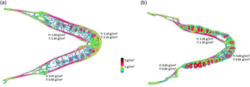
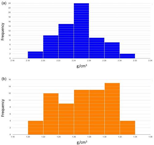
Computed tomography (CT) and microcomputed tomography (μCT) require calibration against density phantoms scanned with specimens or during routine internal calibration for assessment of mineral concentration (MC) and density. In clinical studies involving bone, alternative calibration methods using bodily tissues and fluids (“phantomless” calibration) have been suggested. However, such tissues are seldom available in archeological and osteological research. This study investigates the potential of dental tissue as internal reference for calibration of μCT scans, facilitating the analysis of bone MC. We analyzed 70 molars from 24 extant primate species, including eight human teeth, each scanned with density phantoms for calibration. Our findings indicate that sampling specific regions of molars (lateral aspects of the mesial cusps) yields low variation in enamel and dentine MC values, averaging 1.27 g/cm3 (±0.03) for dentine and 2.25 g/cm3 (±0.03) for enamel. No significant differences were observed across molar types or among scanning procedures, including scanner model, resolution, and filters. An ad hoc test on 12 mandibles revealed low variance in MC between the conventional phantom and dental tissue calibration methods; all 36 measurements (low, medium, and high MC for each mandible) were within 0.05 g/cm3 of each other —81% were within 0.03 g/cm3 and 94% within 0.04 g/cm3. Based on these results, we propose a new “phantomless” calibration technique using these mean enamel and dentine MC values. The presented phantomless calibration method could aid in the assessment of bone pathology and enhance the scope of studies investigating bone structure and physical property variations in archeological, osteological, and laboratory-based research.

 求助内容:
求助内容: 应助结果提醒方式:


