Aditi A Shirke, Jing Wang, Gopolakrishnan Ramamurthy, Arpan Mahanty, Ethan Walker, Lifang Zhang, Abhiram Panigrahi, Xinning Wang, James P Basilion
{"title":"前列腺特异性膜抗原在合成乳腺癌小鼠模型中的表达","authors":"Aditi A Shirke, Jing Wang, Gopolakrishnan Ramamurthy, Arpan Mahanty, Ethan Walker, Lifang Zhang, Abhiram Panigrahi, Xinning Wang, James P Basilion","doi":"10.1007/s11307-024-01920-2","DOIUrl":null,"url":null,"abstract":"<p><strong>Purpose: </strong>Prostate specific membrane antigen (PSMA) has been studied in human breast cancer (BCa) biopsies, however, lack of data on PSMA expression in mouse models impedes development of PSMA-targeted therapies, particularly in improving breast conserving surgery (BCS) margins. This study aimed to validate and characterize the expression of PSMA in murine BCa models, demonstrating that PSMA can be utilized to improve therapies and imaging techniques.</p><p><strong>Methods: </strong>Murine triple negative breast cancer 4T1 cells, and human cell lines, MDA-MB-231, MDA-MB-468, implanted into the mammary fat pads of BALB/c mice, were imaged by our PSMA targeted theranostic agent, PSMA-1-Pc413, and tumor to background ratios (TBR) were calculated to validate selective uptake. Immunohistochemistry was used to correlate PSMA expression in relation to CD31, an endothelial cell biomarker highlighting neovasculature. PSMA expression was also quantified by Reverse Transcriptase Polymerase Chain Reaction (RT-PCR).</p><p><strong>Results: </strong>Accumulation of PSMA-1-Pc413 was observed in 4T1 primary tumors and associated metastases. Average TBR of 4T1 tumors were calculated to be greater than 1.5-ratio at which tumor tissues can be distinguished from normal structures-at peak accumulation with the signal intensity in 4T1 tumors comparable to that in high PSMA expressing PC3-pip tumors. Extraction of 4T1 tumors and lung metastases followed by RT-PCR analysis and PSMA-CD31 co-staining shows that PSMA is consistently localized on tumor neovasculature with no expression in tumor cells and surrounding normal tissues.</p><p><strong>Conclusion: </strong>The selective uptake of PSMA-1-Pc413 in these cancer tissues as well as the characterization and validation of PSMA expression on neovasculature in this syngeneic 4T1 model emphasizes their potential for advancements in targeted therapies and imaging techniques for BCa. PSMA holds great promise as an oncogenic target for BCa and its associated metastases.</p>","PeriodicalId":18760,"journal":{"name":"Molecular Imaging and Biology","volume":" ","pages":"714-728"},"PeriodicalIF":3.0000,"publicationDate":"2024-08-01","publicationTypes":"Journal Article","fieldsOfStudy":null,"isOpenAccess":false,"openAccessPdf":"https://www.ncbi.nlm.nih.gov/pmc/articles/PMC11281974/pdf/","citationCount":"0","resultStr":"{\"title\":\"Prostate Specific Membrane Antigen Expression in a Syngeneic Breast Cancer Mouse Model.\",\"authors\":\"Aditi A Shirke, Jing Wang, Gopolakrishnan Ramamurthy, Arpan Mahanty, Ethan Walker, Lifang Zhang, Abhiram Panigrahi, Xinning Wang, James P Basilion\",\"doi\":\"10.1007/s11307-024-01920-2\",\"DOIUrl\":null,\"url\":null,\"abstract\":\"<p><strong>Purpose: </strong>Prostate specific membrane antigen (PSMA) has been studied in human breast cancer (BCa) biopsies, however, lack of data on PSMA expression in mouse models impedes development of PSMA-targeted therapies, particularly in improving breast conserving surgery (BCS) margins. This study aimed to validate and characterize the expression of PSMA in murine BCa models, demonstrating that PSMA can be utilized to improve therapies and imaging techniques.</p><p><strong>Methods: </strong>Murine triple negative breast cancer 4T1 cells, and human cell lines, MDA-MB-231, MDA-MB-468, implanted into the mammary fat pads of BALB/c mice, were imaged by our PSMA targeted theranostic agent, PSMA-1-Pc413, and tumor to background ratios (TBR) were calculated to validate selective uptake. Immunohistochemistry was used to correlate PSMA expression in relation to CD31, an endothelial cell biomarker highlighting neovasculature. PSMA expression was also quantified by Reverse Transcriptase Polymerase Chain Reaction (RT-PCR).</p><p><strong>Results: </strong>Accumulation of PSMA-1-Pc413 was observed in 4T1 primary tumors and associated metastases. Average TBR of 4T1 tumors were calculated to be greater than 1.5-ratio at which tumor tissues can be distinguished from normal structures-at peak accumulation with the signal intensity in 4T1 tumors comparable to that in high PSMA expressing PC3-pip tumors. Extraction of 4T1 tumors and lung metastases followed by RT-PCR analysis and PSMA-CD31 co-staining shows that PSMA is consistently localized on tumor neovasculature with no expression in tumor cells and surrounding normal tissues.</p><p><strong>Conclusion: </strong>The selective uptake of PSMA-1-Pc413 in these cancer tissues as well as the characterization and validation of PSMA expression on neovasculature in this syngeneic 4T1 model emphasizes their potential for advancements in targeted therapies and imaging techniques for BCa. PSMA holds great promise as an oncogenic target for BCa and its associated metastases.</p>\",\"PeriodicalId\":18760,\"journal\":{\"name\":\"Molecular Imaging and Biology\",\"volume\":\" \",\"pages\":\"714-728\"},\"PeriodicalIF\":3.0000,\"publicationDate\":\"2024-08-01\",\"publicationTypes\":\"Journal Article\",\"fieldsOfStudy\":null,\"isOpenAccess\":false,\"openAccessPdf\":\"https://www.ncbi.nlm.nih.gov/pmc/articles/PMC11281974/pdf/\",\"citationCount\":\"0\",\"resultStr\":null,\"platform\":\"Semanticscholar\",\"paperid\":null,\"PeriodicalName\":\"Molecular Imaging and Biology\",\"FirstCategoryId\":\"3\",\"ListUrlMain\":\"https://doi.org/10.1007/s11307-024-01920-2\",\"RegionNum\":4,\"RegionCategory\":\"医学\",\"ArticlePicture\":[],\"TitleCN\":null,\"AbstractTextCN\":null,\"PMCID\":null,\"EPubDate\":\"2024/5/17 0:00:00\",\"PubModel\":\"Epub\",\"JCR\":\"Q2\",\"JCRName\":\"RADIOLOGY, NUCLEAR MEDICINE & MEDICAL IMAGING\",\"Score\":null,\"Total\":0}","platform":"Semanticscholar","paperid":null,"PeriodicalName":"Molecular Imaging and Biology","FirstCategoryId":"3","ListUrlMain":"https://doi.org/10.1007/s11307-024-01920-2","RegionNum":4,"RegionCategory":"医学","ArticlePicture":[],"TitleCN":null,"AbstractTextCN":null,"PMCID":null,"EPubDate":"2024/5/17 0:00:00","PubModel":"Epub","JCR":"Q2","JCRName":"RADIOLOGY, NUCLEAR MEDICINE & MEDICAL IMAGING","Score":null,"Total":0}
Prostate Specific Membrane Antigen Expression in a Syngeneic Breast Cancer Mouse Model.
Purpose: Prostate specific membrane antigen (PSMA) has been studied in human breast cancer (BCa) biopsies, however, lack of data on PSMA expression in mouse models impedes development of PSMA-targeted therapies, particularly in improving breast conserving surgery (BCS) margins. This study aimed to validate and characterize the expression of PSMA in murine BCa models, demonstrating that PSMA can be utilized to improve therapies and imaging techniques.
Methods: Murine triple negative breast cancer 4T1 cells, and human cell lines, MDA-MB-231, MDA-MB-468, implanted into the mammary fat pads of BALB/c mice, were imaged by our PSMA targeted theranostic agent, PSMA-1-Pc413, and tumor to background ratios (TBR) were calculated to validate selective uptake. Immunohistochemistry was used to correlate PSMA expression in relation to CD31, an endothelial cell biomarker highlighting neovasculature. PSMA expression was also quantified by Reverse Transcriptase Polymerase Chain Reaction (RT-PCR).
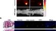
Results: Accumulation of PSMA-1-Pc413 was observed in 4T1 primary tumors and associated metastases. Average TBR of 4T1 tumors were calculated to be greater than 1.5-ratio at which tumor tissues can be distinguished from normal structures-at peak accumulation with the signal intensity in 4T1 tumors comparable to that in high PSMA expressing PC3-pip tumors. Extraction of 4T1 tumors and lung metastases followed by RT-PCR analysis and PSMA-CD31 co-staining shows that PSMA is consistently localized on tumor neovasculature with no expression in tumor cells and surrounding normal tissues.
Conclusion: The selective uptake of PSMA-1-Pc413 in these cancer tissues as well as the characterization and validation of PSMA expression on neovasculature in this syngeneic 4T1 model emphasizes their potential for advancements in targeted therapies and imaging techniques for BCa. PSMA holds great promise as an oncogenic target for BCa and its associated metastases.
期刊介绍:
Molecular Imaging and Biology (MIB) invites original contributions (research articles, review articles, commentaries, etc.) on the utilization of molecular imaging (i.e., nuclear imaging, optical imaging, autoradiography and pathology, MRI, MPI, ultrasound imaging, radiomics/genomics etc.) to investigate questions related to biology and health. The objective of MIB is to provide a forum to the discovery of molecular mechanisms of disease through the use of imaging techniques. We aim to investigate the biological nature of disease in patients and establish new molecular imaging diagnostic and therapy procedures.
Some areas that are covered are:
Preclinical and clinical imaging of macromolecular targets (e.g., genes, receptors, enzymes) involved in significant biological processes.
The design, characterization, and study of new molecular imaging probes and contrast agents for the functional interrogation of macromolecular targets.
Development and evaluation of imaging systems including instrumentation, image reconstruction algorithms, image analysis, and display.
Development of molecular assay approaches leading to quantification of the biological information obtained in molecular imaging.
Study of in vivo animal models of disease for the development of new molecular diagnostics and therapeutics.
Extension of in vitro and in vivo discoveries using disease models, into well designed clinical research investigations.
Clinical molecular imaging involving clinical investigations, clinical trials and medical management or cost-effectiveness studies.

 求助内容:
求助内容: 应助结果提醒方式:


