Owen Paetkau, Sarah Weppler, Jaime Kwok, Harvey C Quon, Wendy Smith, Ekaterina Tchistiakova, Charles Kirkby
{"title":"头颈部放疗中的吞咽困难:咽部解剖和剂量测定的影响。","authors":"Owen Paetkau, Sarah Weppler, Jaime Kwok, Harvey C Quon, Wendy Smith, Ekaterina Tchistiakova, Charles Kirkby","doi":"10.1007/s00455-024-10705-2","DOIUrl":null,"url":null,"abstract":"<p><p>The goal of this study was to identify which anatomical and dosimetric changes correlated with late patient-reported dysphagia throughout the course of head and neck chemo-radiotherapy treatment. The patient cohort (n = 64) considered oropharyngeal and nasopharyngeal patients treated with curative intent, exhibiting no baseline dysphagia with a follow-up time greater than one year. Patients completed the MD Anderson Dysphagia Inventory during a follow-up visit. A composite score was measured ranging from 20 to 100, with a low score indicating a high symptom burden; a score ≤60 indicated patient-reported dysphagia. The pharyngeal (PCM) and cricopharyngeal constrictor muscles (CPM) were contoured on a planning CT image and adapted to weekly cone-beam CT anatomy using deformable image registration and dose was accumulated using weighted dose-volume histogram curves. The PCM and CPM were examined for volume, thickness, and dosimetric changes across treatment with the results correlated to symptom group. Anatomical evaluation indicated the PCM thickness increased more during treatment for patients with dysphagia, with base of C2 vertebrae (p = 0.04) and superior-inferior middle PCM (p = 0.01) thicknesses indicating a 1.0-1.5 mm increase. The planned and delivered mean dose and DVH metrics to PCM and CPM were found to be within random error measured for the dose accumulation, indicating delivered and planned dose are equivalent. The PCM and CPM organs were found to lie approximately 5 mm closer to high dose gradients in patients exhibiting dysphagia. The volume, thickness, and high dose gradient metrics may be useful metrics to identify patients at risk of late patient-reported dysphagia.</p>","PeriodicalId":11508,"journal":{"name":"Dysphagia","volume":" ","pages":"77-87"},"PeriodicalIF":2.2000,"publicationDate":"2025-02-01","publicationTypes":"Journal Article","fieldsOfStudy":null,"isOpenAccess":false,"openAccessPdf":"","citationCount":"0","resultStr":"{\"title\":\"Dysphagia in Head and Neck Radiotherapy: The Influence of Pharyngeal Constrictor Anatomy and Dosimetry.\",\"authors\":\"Owen Paetkau, Sarah Weppler, Jaime Kwok, Harvey C Quon, Wendy Smith, Ekaterina Tchistiakova, Charles Kirkby\",\"doi\":\"10.1007/s00455-024-10705-2\",\"DOIUrl\":null,\"url\":null,\"abstract\":\"<p><p>The goal of this study was to identify which anatomical and dosimetric changes correlated with late patient-reported dysphagia throughout the course of head and neck chemo-radiotherapy treatment. The patient cohort (n = 64) considered oropharyngeal and nasopharyngeal patients treated with curative intent, exhibiting no baseline dysphagia with a follow-up time greater than one year. Patients completed the MD Anderson Dysphagia Inventory during a follow-up visit. A composite score was measured ranging from 20 to 100, with a low score indicating a high symptom burden; a score ≤60 indicated patient-reported dysphagia. The pharyngeal (PCM) and cricopharyngeal constrictor muscles (CPM) were contoured on a planning CT image and adapted to weekly cone-beam CT anatomy using deformable image registration and dose was accumulated using weighted dose-volume histogram curves. The PCM and CPM were examined for volume, thickness, and dosimetric changes across treatment with the results correlated to symptom group. Anatomical evaluation indicated the PCM thickness increased more during treatment for patients with dysphagia, with base of C2 vertebrae (p = 0.04) and superior-inferior middle PCM (p = 0.01) thicknesses indicating a 1.0-1.5 mm increase. The planned and delivered mean dose and DVH metrics to PCM and CPM were found to be within random error measured for the dose accumulation, indicating delivered and planned dose are equivalent. The PCM and CPM organs were found to lie approximately 5 mm closer to high dose gradients in patients exhibiting dysphagia. The volume, thickness, and high dose gradient metrics may be useful metrics to identify patients at risk of late patient-reported dysphagia.</p>\",\"PeriodicalId\":11508,\"journal\":{\"name\":\"Dysphagia\",\"volume\":\" \",\"pages\":\"77-87\"},\"PeriodicalIF\":2.2000,\"publicationDate\":\"2025-02-01\",\"publicationTypes\":\"Journal Article\",\"fieldsOfStudy\":null,\"isOpenAccess\":false,\"openAccessPdf\":\"\",\"citationCount\":\"0\",\"resultStr\":null,\"platform\":\"Semanticscholar\",\"paperid\":null,\"PeriodicalName\":\"Dysphagia\",\"FirstCategoryId\":\"3\",\"ListUrlMain\":\"https://doi.org/10.1007/s00455-024-10705-2\",\"RegionNum\":3,\"RegionCategory\":\"医学\",\"ArticlePicture\":[],\"TitleCN\":null,\"AbstractTextCN\":null,\"PMCID\":null,\"EPubDate\":\"2024/5/16 0:00:00\",\"PubModel\":\"Epub\",\"JCR\":\"Q1\",\"JCRName\":\"OTORHINOLARYNGOLOGY\",\"Score\":null,\"Total\":0}","platform":"Semanticscholar","paperid":null,"PeriodicalName":"Dysphagia","FirstCategoryId":"3","ListUrlMain":"https://doi.org/10.1007/s00455-024-10705-2","RegionNum":3,"RegionCategory":"医学","ArticlePicture":[],"TitleCN":null,"AbstractTextCN":null,"PMCID":null,"EPubDate":"2024/5/16 0:00:00","PubModel":"Epub","JCR":"Q1","JCRName":"OTORHINOLARYNGOLOGY","Score":null,"Total":0}
Dysphagia in Head and Neck Radiotherapy: The Influence of Pharyngeal Constrictor Anatomy and Dosimetry.
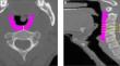
The goal of this study was to identify which anatomical and dosimetric changes correlated with late patient-reported dysphagia throughout the course of head and neck chemo-radiotherapy treatment. The patient cohort (n = 64) considered oropharyngeal and nasopharyngeal patients treated with curative intent, exhibiting no baseline dysphagia with a follow-up time greater than one year. Patients completed the MD Anderson Dysphagia Inventory during a follow-up visit. A composite score was measured ranging from 20 to 100, with a low score indicating a high symptom burden; a score ≤60 indicated patient-reported dysphagia. The pharyngeal (PCM) and cricopharyngeal constrictor muscles (CPM) were contoured on a planning CT image and adapted to weekly cone-beam CT anatomy using deformable image registration and dose was accumulated using weighted dose-volume histogram curves. The PCM and CPM were examined for volume, thickness, and dosimetric changes across treatment with the results correlated to symptom group. Anatomical evaluation indicated the PCM thickness increased more during treatment for patients with dysphagia, with base of C2 vertebrae (p = 0.04) and superior-inferior middle PCM (p = 0.01) thicknesses indicating a 1.0-1.5 mm increase. The planned and delivered mean dose and DVH metrics to PCM and CPM were found to be within random error measured for the dose accumulation, indicating delivered and planned dose are equivalent. The PCM and CPM organs were found to lie approximately 5 mm closer to high dose gradients in patients exhibiting dysphagia. The volume, thickness, and high dose gradient metrics may be useful metrics to identify patients at risk of late patient-reported dysphagia.
期刊介绍:
Dysphagia aims to serve as a voice for the benefit of the patient. The journal is devoted exclusively to swallowing and its disorders. The purpose of the journal is to provide a source of information to the flourishing dysphagia community. Over the past years, the field of dysphagia has grown rapidly, and the community of dysphagia researchers have galvanized with ambition to represent dysphagia patients. In addition to covering a myriad of disciplines in medicine and speech pathology, the following topics are also covered, but are not limited to: bio-engineering, deglutition, esophageal motility, immunology, and neuro-gastroenterology. The journal aims to foster a growing need for further dysphagia investigation, to disseminate knowledge through research, and to stimulate communication among interested professionals. The journal publishes original papers, technical and instrumental notes, letters to the editor, and review articles.
 求助内容:
求助内容: 应助结果提醒方式:


