Nils D Forkert, Sarah J MacEachern, Allison K Duh, Peter Moon, Sarah Lee, Kristen W Yeom
{"title":"先天性心脏病患儿的深层灰质结构发生变化。","authors":"Nils D Forkert, Sarah J MacEachern, Allison K Duh, Peter Moon, Sarah Lee, Kristen W Yeom","doi":"10.1007/s00062-024-01417-z","DOIUrl":null,"url":null,"abstract":"<p><strong>Background and objectives: </strong>Children with congenital heart diseases (CHDs) have an increased risk of developing neurologic deficits, even in the absence of apparent brain pathology. The aim of this work was to compare quantitative macro- and microstructural properties of subcortical gray matter structures of pediatric CHD patients with normal appearing brain magnetic resonance imaging to healthy controls.</p><p><strong>Methods: </strong>We retrospectively reviewed children with coarctation of the aorta (COA) and hypoplastic left heart syndrome (HLHS) admitted to our hospital. We identified 24 pediatric CHD patients (17 COA, 7 HLHS) with normal-appearing brain MRI. Using an atlas-based approach, the volume and apparent diffusion coefficient (ADC) were determined for the thalamus, caudate, putamen, pallidum, hippocampus, amygdala, nucleus accumbens, cerebral white matter, cerebral cortex, and brainstem. Multivariate statistics were used to compare the extracted values to reference values from 100 typically developing children without any known cardiac or neurological diseases.</p><p><strong>Results: </strong>Multivariate analysis of covariance using the regional ADC and volume values as dependent variables and age and sex as co-variates revealed a significant difference between pediatric CHD patients and healthy controls (p < 0.001). Post-hoc comparisons demonstrated significantly reduced brain volumes in most subcortical brain regions investigated and elevated ADC values in the thalamus for children with CHD. No significant differences were found comparing children with COA and HLHS.</p><p><strong>Conclusions: </strong>Despite normal appearing brain MRI, children with CHD exhibit wide-spread macro-structural and regional micro-structural differences of subcortical brain structures compared to healthy controls, which could negatively impact neurodevelopment, leading to neurological deficits in childhood and beyond.</p>","PeriodicalId":49298,"journal":{"name":"Clinical Neuroradiology","volume":" ","pages":"771-778"},"PeriodicalIF":2.6000,"publicationDate":"2024-12-01","publicationTypes":"Journal Article","fieldsOfStudy":null,"isOpenAccess":false,"openAccessPdf":"","citationCount":"0","resultStr":"{\"title\":\"Children with Congenital Heart Diseases Exhibit Altered Deep Gray Matter Structures.\",\"authors\":\"Nils D Forkert, Sarah J MacEachern, Allison K Duh, Peter Moon, Sarah Lee, Kristen W Yeom\",\"doi\":\"10.1007/s00062-024-01417-z\",\"DOIUrl\":null,\"url\":null,\"abstract\":\"<p><strong>Background and objectives: </strong>Children with congenital heart diseases (CHDs) have an increased risk of developing neurologic deficits, even in the absence of apparent brain pathology. The aim of this work was to compare quantitative macro- and microstructural properties of subcortical gray matter structures of pediatric CHD patients with normal appearing brain magnetic resonance imaging to healthy controls.</p><p><strong>Methods: </strong>We retrospectively reviewed children with coarctation of the aorta (COA) and hypoplastic left heart syndrome (HLHS) admitted to our hospital. We identified 24 pediatric CHD patients (17 COA, 7 HLHS) with normal-appearing brain MRI. Using an atlas-based approach, the volume and apparent diffusion coefficient (ADC) were determined for the thalamus, caudate, putamen, pallidum, hippocampus, amygdala, nucleus accumbens, cerebral white matter, cerebral cortex, and brainstem. Multivariate statistics were used to compare the extracted values to reference values from 100 typically developing children without any known cardiac or neurological diseases.</p><p><strong>Results: </strong>Multivariate analysis of covariance using the regional ADC and volume values as dependent variables and age and sex as co-variates revealed a significant difference between pediatric CHD patients and healthy controls (p < 0.001). Post-hoc comparisons demonstrated significantly reduced brain volumes in most subcortical brain regions investigated and elevated ADC values in the thalamus for children with CHD. No significant differences were found comparing children with COA and HLHS.</p><p><strong>Conclusions: </strong>Despite normal appearing brain MRI, children with CHD exhibit wide-spread macro-structural and regional micro-structural differences of subcortical brain structures compared to healthy controls, which could negatively impact neurodevelopment, leading to neurological deficits in childhood and beyond.</p>\",\"PeriodicalId\":49298,\"journal\":{\"name\":\"Clinical Neuroradiology\",\"volume\":\" \",\"pages\":\"771-778\"},\"PeriodicalIF\":2.6000,\"publicationDate\":\"2024-12-01\",\"publicationTypes\":\"Journal Article\",\"fieldsOfStudy\":null,\"isOpenAccess\":false,\"openAccessPdf\":\"\",\"citationCount\":\"0\",\"resultStr\":null,\"platform\":\"Semanticscholar\",\"paperid\":null,\"PeriodicalName\":\"Clinical Neuroradiology\",\"FirstCategoryId\":\"1085\",\"ListUrlMain\":\"https://doi.org/10.1007/s00062-024-01417-z\",\"RegionNum\":3,\"RegionCategory\":\"医学\",\"ArticlePicture\":[],\"TitleCN\":null,\"AbstractTextCN\":null,\"PMCID\":null,\"EPubDate\":\"2024/5/14 0:00:00\",\"PubModel\":\"Epub\",\"JCR\":\"Q2\",\"JCRName\":\"CLINICAL NEUROLOGY\",\"Score\":null,\"Total\":0}","platform":"Semanticscholar","paperid":null,"PeriodicalName":"Clinical Neuroradiology","FirstCategoryId":"1085","ListUrlMain":"https://doi.org/10.1007/s00062-024-01417-z","RegionNum":3,"RegionCategory":"医学","ArticlePicture":[],"TitleCN":null,"AbstractTextCN":null,"PMCID":null,"EPubDate":"2024/5/14 0:00:00","PubModel":"Epub","JCR":"Q2","JCRName":"CLINICAL NEUROLOGY","Score":null,"Total":0}
Children with Congenital Heart Diseases Exhibit Altered Deep Gray Matter Structures.
Background and objectives: Children with congenital heart diseases (CHDs) have an increased risk of developing neurologic deficits, even in the absence of apparent brain pathology. The aim of this work was to compare quantitative macro- and microstructural properties of subcortical gray matter structures of pediatric CHD patients with normal appearing brain magnetic resonance imaging to healthy controls.
Methods: We retrospectively reviewed children with coarctation of the aorta (COA) and hypoplastic left heart syndrome (HLHS) admitted to our hospital. We identified 24 pediatric CHD patients (17 COA, 7 HLHS) with normal-appearing brain MRI. Using an atlas-based approach, the volume and apparent diffusion coefficient (ADC) were determined for the thalamus, caudate, putamen, pallidum, hippocampus, amygdala, nucleus accumbens, cerebral white matter, cerebral cortex, and brainstem. Multivariate statistics were used to compare the extracted values to reference values from 100 typically developing children without any known cardiac or neurological diseases.
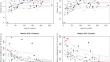
Results: Multivariate analysis of covariance using the regional ADC and volume values as dependent variables and age and sex as co-variates revealed a significant difference between pediatric CHD patients and healthy controls (p < 0.001). Post-hoc comparisons demonstrated significantly reduced brain volumes in most subcortical brain regions investigated and elevated ADC values in the thalamus for children with CHD. No significant differences were found comparing children with COA and HLHS.
Conclusions: Despite normal appearing brain MRI, children with CHD exhibit wide-spread macro-structural and regional micro-structural differences of subcortical brain structures compared to healthy controls, which could negatively impact neurodevelopment, leading to neurological deficits in childhood and beyond.
期刊介绍:
Clinical Neuroradiology provides current information, original contributions, and reviews in the field of neuroradiology. An interdisciplinary approach is accomplished by diagnostic and therapeutic contributions related to associated subjects.
The international coverage and relevance of the journal is underlined by its being the official journal of the German, Swiss, and Austrian Societies of Neuroradiology.
 求助内容:
求助内容: 应助结果提醒方式:


