Dimitrios Goutas, Pauline Hayenne, Franck Tirode, Daniel Pissaloux, Iwei Yeh, Arnaud de la Fouchardiere
{"title":"蛋白激酶 C 融合蓝痣中的平滑肌增生:12 个病例的报告。","authors":"Dimitrios Goutas, Pauline Hayenne, Franck Tirode, Daniel Pissaloux, Iwei Yeh, Arnaud de la Fouchardiere","doi":"10.1111/his.15211","DOIUrl":null,"url":null,"abstract":"<div>\n \n <section>\n \n <h3> Background and aims</h3>\n \n <p>PKC-fused blue naevi are a recently described group of melanocytic tumours that have distinctive morphological features, including a pigmented epithelioid melanocytoma-like junctional component or a dermal biphasic architecture associating with naevocytoid nests surrounded by dendritic and spindled pigmented melanocytes (so-called ‘combined common and blue naevus’). There have been reports of smooth muscle hyperplasia in a hamartoma-like pattern in cases of combined blue naevi without genetic exploration.</p>\n </section>\n \n <section>\n \n <h3> Materials and methods</h3>\n \n <p>Herein, we describe 12 cases of protein kinase C (PKC)-fused blue tumours associated with a co-existing smooth muscle hyperplasia, identified from a total of 98 PKC-fused melanocytic tumours. Archived slides of PKC-fused blue naevi with haematoxylin, eosin and phloxin staining, immunohistochemistry and molecular confirmation of a PKC-fusion by fluorescence <i>in-situ</i> hybridisation (FISH) or RNAseq were re-evaluated for identification of notable smooth muscle hyperplasia. Fifty-one of these slides had already been studied in a previous publication from our group.</p>\n </section>\n \n <section>\n \n <h3> Results</h3>\n \n <p>The hyperplasia ranged from hypertrophic arrector pili muscles to extensive horizontal bundles of disorganised fibres constantly associated and limited within a biphasic dermal melanocytic component. At least one arrector pili muscle was always visible within the tumour, with occasionally direct extension of the hyperplastic fibres from the main muscle body. These muscle fibres were devoid of a PKC-fusion signal by FISH. PKC molecules are involved in the regulation of smooth muscle function, offering an explanatory framework.</p>\n </section>\n \n <section>\n \n <h3> Conclusions</h3>\n \n <p>These data suggest incorporating smooth muscle hyperplasia as a diagnostic morphological feature of PKC-fused blue melanocytic tumours.</p>\n </section>\n </div>","PeriodicalId":13219,"journal":{"name":"Histopathology","volume":null,"pages":null},"PeriodicalIF":3.9000,"publicationDate":"2024-05-15","publicationTypes":"Journal Article","fieldsOfStudy":null,"isOpenAccess":false,"openAccessPdf":"","citationCount":"0","resultStr":"{\"title\":\"Smooth muscle hyperplasia in protein kinase C-fused blue naevi: Report of 12 cases\",\"authors\":\"Dimitrios Goutas, Pauline Hayenne, Franck Tirode, Daniel Pissaloux, Iwei Yeh, Arnaud de la Fouchardiere\",\"doi\":\"10.1111/his.15211\",\"DOIUrl\":null,\"url\":null,\"abstract\":\"<div>\\n \\n <section>\\n \\n <h3> Background and aims</h3>\\n \\n <p>PKC-fused blue naevi are a recently described group of melanocytic tumours that have distinctive morphological features, including a pigmented epithelioid melanocytoma-like junctional component or a dermal biphasic architecture associating with naevocytoid nests surrounded by dendritic and spindled pigmented melanocytes (so-called ‘combined common and blue naevus’). There have been reports of smooth muscle hyperplasia in a hamartoma-like pattern in cases of combined blue naevi without genetic exploration.</p>\\n </section>\\n \\n <section>\\n \\n <h3> Materials and methods</h3>\\n \\n <p>Herein, we describe 12 cases of protein kinase C (PKC)-fused blue tumours associated with a co-existing smooth muscle hyperplasia, identified from a total of 98 PKC-fused melanocytic tumours. Archived slides of PKC-fused blue naevi with haematoxylin, eosin and phloxin staining, immunohistochemistry and molecular confirmation of a PKC-fusion by fluorescence <i>in-situ</i> hybridisation (FISH) or RNAseq were re-evaluated for identification of notable smooth muscle hyperplasia. Fifty-one of these slides had already been studied in a previous publication from our group.</p>\\n </section>\\n \\n <section>\\n \\n <h3> Results</h3>\\n \\n <p>The hyperplasia ranged from hypertrophic arrector pili muscles to extensive horizontal bundles of disorganised fibres constantly associated and limited within a biphasic dermal melanocytic component. At least one arrector pili muscle was always visible within the tumour, with occasionally direct extension of the hyperplastic fibres from the main muscle body. These muscle fibres were devoid of a PKC-fusion signal by FISH. PKC molecules are involved in the regulation of smooth muscle function, offering an explanatory framework.</p>\\n </section>\\n \\n <section>\\n \\n <h3> Conclusions</h3>\\n \\n <p>These data suggest incorporating smooth muscle hyperplasia as a diagnostic morphological feature of PKC-fused blue melanocytic tumours.</p>\\n </section>\\n </div>\",\"PeriodicalId\":13219,\"journal\":{\"name\":\"Histopathology\",\"volume\":null,\"pages\":null},\"PeriodicalIF\":3.9000,\"publicationDate\":\"2024-05-15\",\"publicationTypes\":\"Journal Article\",\"fieldsOfStudy\":null,\"isOpenAccess\":false,\"openAccessPdf\":\"\",\"citationCount\":\"0\",\"resultStr\":null,\"platform\":\"Semanticscholar\",\"paperid\":null,\"PeriodicalName\":\"Histopathology\",\"FirstCategoryId\":\"3\",\"ListUrlMain\":\"https://onlinelibrary.wiley.com/doi/10.1111/his.15211\",\"RegionNum\":2,\"RegionCategory\":\"医学\",\"ArticlePicture\":[],\"TitleCN\":null,\"AbstractTextCN\":null,\"PMCID\":null,\"EPubDate\":\"\",\"PubModel\":\"\",\"JCR\":\"Q2\",\"JCRName\":\"CELL BIOLOGY\",\"Score\":null,\"Total\":0}","platform":"Semanticscholar","paperid":null,"PeriodicalName":"Histopathology","FirstCategoryId":"3","ListUrlMain":"https://onlinelibrary.wiley.com/doi/10.1111/his.15211","RegionNum":2,"RegionCategory":"医学","ArticlePicture":[],"TitleCN":null,"AbstractTextCN":null,"PMCID":null,"EPubDate":"","PubModel":"","JCR":"Q2","JCRName":"CELL BIOLOGY","Score":null,"Total":0}
Smooth muscle hyperplasia in protein kinase C-fused blue naevi: Report of 12 cases
Background and aims
PKC-fused blue naevi are a recently described group of melanocytic tumours that have distinctive morphological features, including a pigmented epithelioid melanocytoma-like junctional component or a dermal biphasic architecture associating with naevocytoid nests surrounded by dendritic and spindled pigmented melanocytes (so-called ‘combined common and blue naevus’). There have been reports of smooth muscle hyperplasia in a hamartoma-like pattern in cases of combined blue naevi without genetic exploration.
Materials and methods
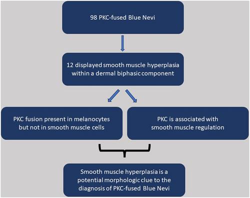
Herein, we describe 12 cases of protein kinase C (PKC)-fused blue tumours associated with a co-existing smooth muscle hyperplasia, identified from a total of 98 PKC-fused melanocytic tumours. Archived slides of PKC-fused blue naevi with haematoxylin, eosin and phloxin staining, immunohistochemistry and molecular confirmation of a PKC-fusion by fluorescence in-situ hybridisation (FISH) or RNAseq were re-evaluated for identification of notable smooth muscle hyperplasia. Fifty-one of these slides had already been studied in a previous publication from our group.
Results
The hyperplasia ranged from hypertrophic arrector pili muscles to extensive horizontal bundles of disorganised fibres constantly associated and limited within a biphasic dermal melanocytic component. At least one arrector pili muscle was always visible within the tumour, with occasionally direct extension of the hyperplastic fibres from the main muscle body. These muscle fibres were devoid of a PKC-fusion signal by FISH. PKC molecules are involved in the regulation of smooth muscle function, offering an explanatory framework.
Conclusions
These data suggest incorporating smooth muscle hyperplasia as a diagnostic morphological feature of PKC-fused blue melanocytic tumours.
期刊介绍:
Histopathology is an international journal intended to be of practical value to surgical and diagnostic histopathologists, and to investigators of human disease who employ histopathological methods. Our primary purpose is to publish advances in pathology, in particular those applicable to clinical practice and contributing to the better understanding of human disease.
 求助内容:
求助内容: 应助结果提醒方式:


